{"title":"Muscarinic Receptor Stimulation Does Not Inhibit Voltage-dependent Ca<sup>2+</sup> Channels in Rat Adrenal Medullary Chromaffin Cells.","authors":"Keita Harada, Masumi Inoue","doi":"10.1267/ahc.23-00042","DOIUrl":null,"url":null,"abstract":"<p><p>Adrenal medullary chromaffin (AMC) and sympathetic ganglion cells are derived from the neural crest and show a similar developmental path. Thus, these two cell types have many common properties in membrane excitability and signaling. However, AMC cells function as endocrine cells while sympathetic ganglion cells are neurons. In rat sympathetic ganglion cells, muscarinic M<sub>1</sub> and M<sub>4</sub> receptors mediate excitation and inhibition via suppression of M-type K<sup>+</sup> channels and suppression of voltage-dependent Ca<sup>2+</sup> channels, respectively. On the other hand, M<sub>1</sub> receptor stimulation in rat AMC cells also produces excitation by suppressing TWIK-related acid sensitive K<sup>+</sup> (TASK) channels. However, whether M<sub>4</sub> receptors are coupled with voltage-dependent Ca<sup>2+</sup> channel suppression is unclear. We explore this issue electrophysiologically and biochemically. Electrical stimulation of nerve fibers in rat adrenal glands trans-synaptically increased the Ca<sup>2+</sup> signal in AMC cells. This electrically evoked increased Ca<sup>2+</sup> signal was not altered during muscarine-induced increase in Ca<sup>2+</sup> signal, whereas it decreased significantly during a GABA-induced increase, due to a shunt effect of increased Cl<sup>-</sup> conductance. The whole-cell current recordings revealed that voltage-dependent Ca<sup>2+</sup> currents in AMC cells were suppressed by adenosine triphosphate, but not by muscarinic agonists. The fractionation analysis and immunocytochemistry indicated that Ca<sub>V</sub>1.2 Ca<sup>2+</sup> channels and M<sub>4</sub> receptors are located in the raft and non-raft membrane domains, respectively. We concluded that muscarinic stimulation in rat AMC cells does not produce voltage-dependent Ca<sup>2+</sup> channel inhibition. This lack of muscarinic inhibition is at least partly due to physical separation of voltage-dependent Ca<sup>2+</sup> channels and M<sub>4</sub> receptors in the plasma membrane.</p>","PeriodicalId":6888,"journal":{"name":"Acta Histochemica Et Cytochemica","volume":"56 4","pages":"67-75"},"PeriodicalIF":1.8000,"publicationDate":"2023-08-30","publicationTypes":"Journal Article","fieldsOfStudy":null,"isOpenAccess":false,"openAccessPdf":"https://ftp.ncbi.nlm.nih.gov/pub/pmc/oa_pdf/7a/6b/ahc-056-67.PMC10480484.pdf","citationCount":"0","resultStr":null,"platform":"Semanticscholar","paperid":null,"PeriodicalName":"Acta Histochemica Et Cytochemica","FirstCategoryId":"99","ListUrlMain":"https://doi.org/10.1267/ahc.23-00042","RegionNum":4,"RegionCategory":"生物学","ArticlePicture":[],"TitleCN":null,"AbstractTextCN":null,"PMCID":null,"EPubDate":"","PubModel":"","JCR":"Q4","JCRName":"CELL BIOLOGY","Score":null,"Total":0}
引用次数: 0
Abstract
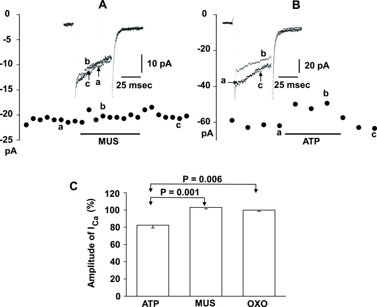
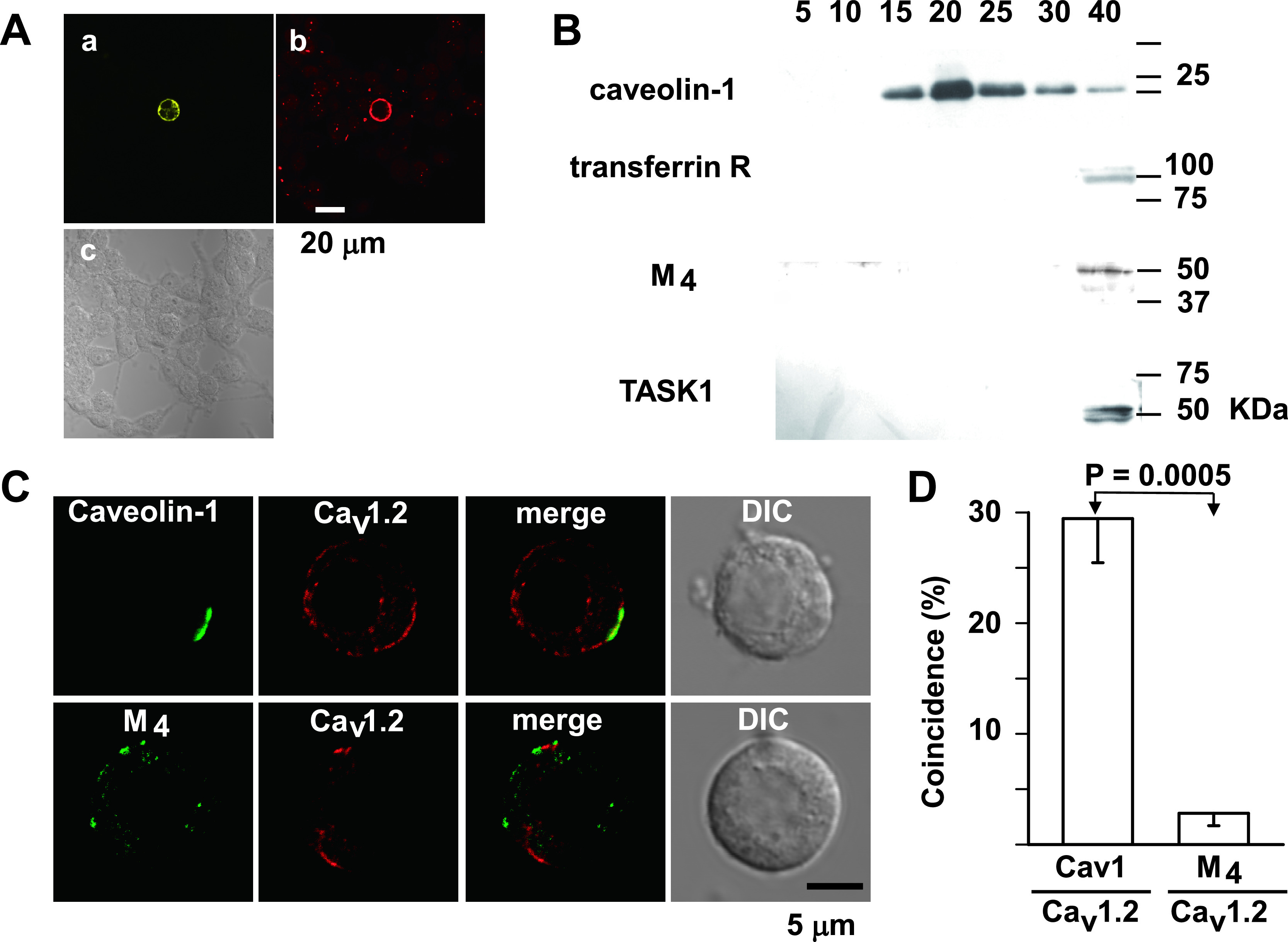
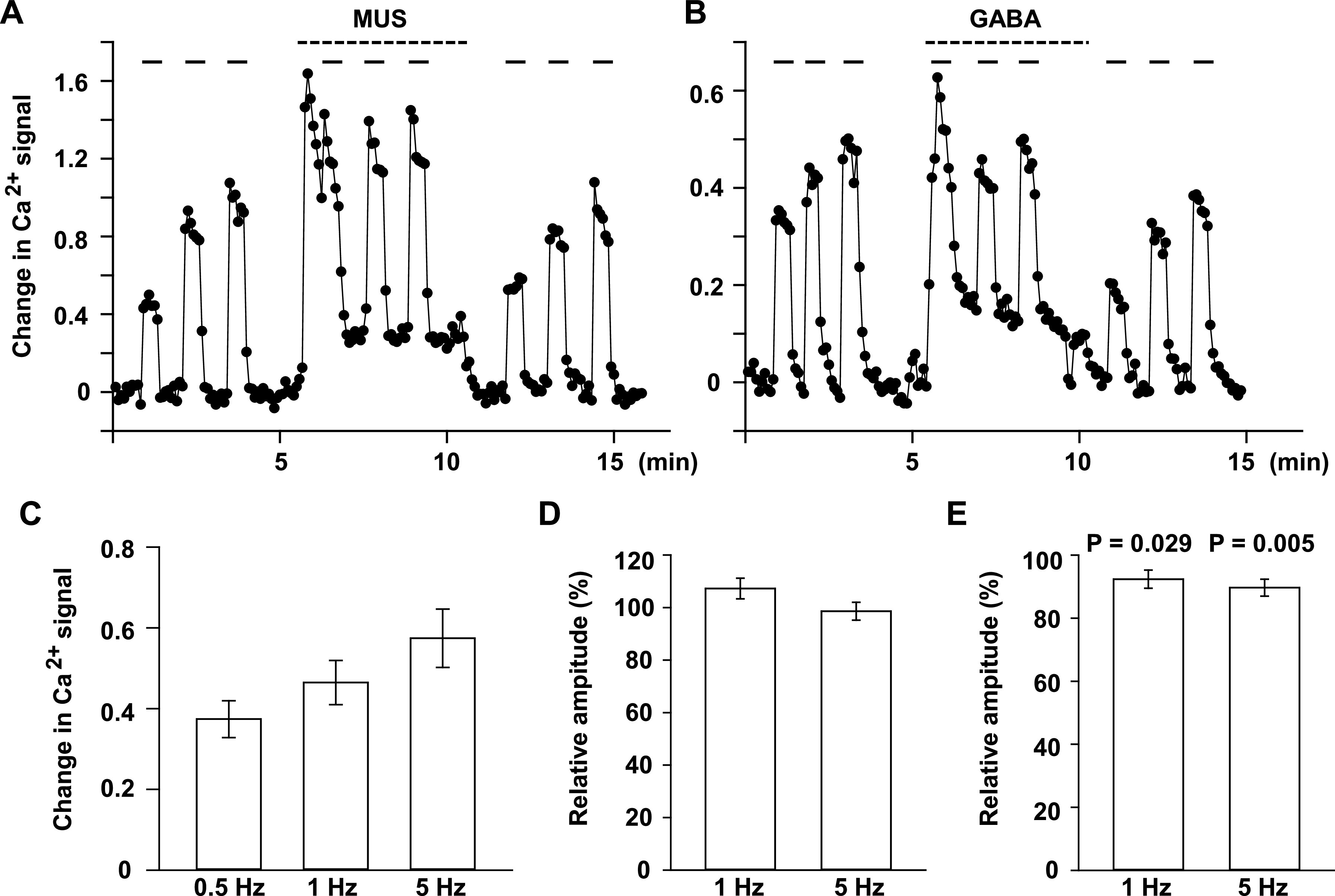
Adrenal medullary chromaffin (AMC) and sympathetic ganglion cells are derived from the neural crest and show a similar developmental path. Thus, these two cell types have many common properties in membrane excitability and signaling. However, AMC cells function as endocrine cells while sympathetic ganglion cells are neurons. In rat sympathetic ganglion cells, muscarinic M1 and M4 receptors mediate excitation and inhibition via suppression of M-type K+ channels and suppression of voltage-dependent Ca2+ channels, respectively. On the other hand, M1 receptor stimulation in rat AMC cells also produces excitation by suppressing TWIK-related acid sensitive K+ (TASK) channels. However, whether M4 receptors are coupled with voltage-dependent Ca2+ channel suppression is unclear. We explore this issue electrophysiologically and biochemically. Electrical stimulation of nerve fibers in rat adrenal glands trans-synaptically increased the Ca2+ signal in AMC cells. This electrically evoked increased Ca2+ signal was not altered during muscarine-induced increase in Ca2+ signal, whereas it decreased significantly during a GABA-induced increase, due to a shunt effect of increased Cl- conductance. The whole-cell current recordings revealed that voltage-dependent Ca2+ currents in AMC cells were suppressed by adenosine triphosphate, but not by muscarinic agonists. The fractionation analysis and immunocytochemistry indicated that CaV1.2 Ca2+ channels and M4 receptors are located in the raft and non-raft membrane domains, respectively. We concluded that muscarinic stimulation in rat AMC cells does not produce voltage-dependent Ca2+ channel inhibition. This lack of muscarinic inhibition is at least partly due to physical separation of voltage-dependent Ca2+ channels and M4 receptors in the plasma membrane.
期刊介绍:
Acta Histochemica et Cytochemica is the official online journal of the Japan Society of Histochemistry and Cytochemistry. It is intended primarily for rapid publication of concise, original articles in the fields of histochemistry and cytochemistry. Manuscripts oriented towards methodological subjects that contain significant technical advances in these fields are also welcome. Manuscripts in English are accepted from investigators in any country, whether or not they are members of the Japan Society of Histochemistry and Cytochemistry. Manuscripts should be original work that has not been previously published and is not being considered for publication elsewhere, with the exception of abstracts. Manuscripts with essentially the same content as a paper that has been published or accepted, or is under consideration for publication, will not be considered. All submitted papers will be peer-reviewed by at least two referees selected by an appropriate Associate Editor. Acceptance is based on scientific significance, originality, and clarity. When required, a revised manuscript should be submitted within 3 months, otherwise it will be considered to be a new submission. The Editor-in-Chief will make all final decisions regarding acceptance.


 分享
分享
 求助内容:
求助内容: 应助结果提醒方式:
 扫码关注我们
