{"title":"Bilateral Cerebral Ptosis in a Patient with Subdural Hemorrhage: a Case Report.","authors":"Ji Hye Kang, Sunha Park, Young-Ah Choi","doi":"10.12786/bn.2021.14.e17","DOIUrl":null,"url":null,"abstract":"<p><p>Although cerebral ptosis is rare, it is commonly associated with unilateral right cerebral hemisphere lesions. We report a case of a 79-year-old woman who presented with bilateral complete ptosis after a traumatic right fronto-temporo-parietal subdural hemorrhage (SDH). Bilateral ptosis was the primary manifestation of the acute right SDH, and the patient had no parenchymal lesion. Her prognosis was good, and she made a complete recovery. Right hemispheric hypoperfusion, as demonstrated on brain perfusion single-photon emission computed tomography, implied that the lateralization of eyelid control was in the right hemisphere, in line with previous reports.</p>","PeriodicalId":72442,"journal":{"name":"Brain & NeuroRehabilitation","volume":"14 2","pages":"e17"},"PeriodicalIF":0.0000,"publicationDate":"2021-07-01","publicationTypes":"Journal Article","fieldsOfStudy":null,"isOpenAccess":false,"openAccessPdf":"https://ftp.ncbi.nlm.nih.gov/pub/pmc/oa_pdf/a4/d3/bn-14-e17.PMC9879497.pdf","citationCount":"0","resultStr":null,"platform":"Semanticscholar","paperid":null,"PeriodicalName":"Brain & NeuroRehabilitation","FirstCategoryId":"1085","ListUrlMain":"https://doi.org/10.12786/bn.2021.14.e17","RegionNum":0,"RegionCategory":null,"ArticlePicture":[],"TitleCN":null,"AbstractTextCN":null,"PMCID":null,"EPubDate":"","PubModel":"","JCR":"","JCRName":"","Score":null,"Total":0}
引用次数: 0
Abstract
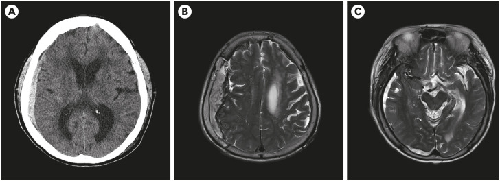
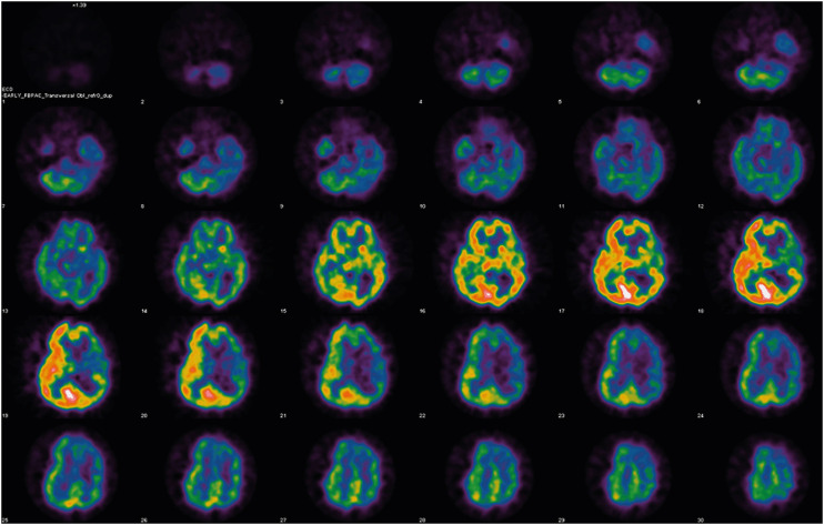
Although cerebral ptosis is rare, it is commonly associated with unilateral right cerebral hemisphere lesions. We report a case of a 79-year-old woman who presented with bilateral complete ptosis after a traumatic right fronto-temporo-parietal subdural hemorrhage (SDH). Bilateral ptosis was the primary manifestation of the acute right SDH, and the patient had no parenchymal lesion. Her prognosis was good, and she made a complete recovery. Right hemispheric hypoperfusion, as demonstrated on brain perfusion single-photon emission computed tomography, implied that the lateralization of eyelid control was in the right hemisphere, in line with previous reports.


 分享
分享

 求助内容:
求助内容: 应助结果提醒方式:
 扫码关注我们
