{"title":"Commentary: locating the restriction point.","authors":"Robert F Brooks","doi":"10.1186/s13008-023-00085-8","DOIUrl":null,"url":null,"abstract":"<p><p>Attempts to map the Restriction Point in the mammalian cell cycle typically involve stimulating quiescent cells with mitogens for increasing intervals, removing the stimulus and then determining the proportion of cells that reach S phase at some point later. This \"fixed point\" estimate assumes that further cell cycle commitment ceases as soon as the stimulus is removed. In fact, kinetic analysis shows that the probability of cell cycle commitment does not fall back to its initial low value, immediately after a pulse of mitogens, but may instead remain slightly elevated for some while afterwards, compared to the starting quiescent population. Thus, cells entering S phase after a brief exposure to mitogens are not those that pass the Restriction Point early. Rather, they represent cells that continue on to S phase as a result of this residual, low probability of cell cycle commitment. Instead, the mitogen-regulated process(es) affecting the probability of cell cycle commitment are much closer to the start of S phase itself. Since the acquisition of (apparent) mitogen independence is such a poor indicator of the timing of cell cycle commitment, it is argued that a better measure is the point of insensitivity to CDK4,6 inhibitors such as palbociclib, which indicates when hyperphosphorylation of the Retinoblastoma Protein, RB, ceases to be dependent on mitogen-signalling pathways regulating CDK4,6/cyclin D activity.</p>","PeriodicalId":49263,"journal":{"name":"Cell Division","volume":"18 1","pages":"2"},"PeriodicalIF":2.2000,"publicationDate":"2023-02-10","publicationTypes":"Journal Article","fieldsOfStudy":null,"isOpenAccess":false,"openAccessPdf":"https://www.ncbi.nlm.nih.gov/pmc/articles/PMC9912616/pdf/","citationCount":"2","resultStr":null,"platform":"Semanticscholar","paperid":null,"PeriodicalName":"Cell Division","FirstCategoryId":"99","ListUrlMain":"https://doi.org/10.1186/s13008-023-00085-8","RegionNum":4,"RegionCategory":"生物学","ArticlePicture":[],"TitleCN":null,"AbstractTextCN":null,"PMCID":null,"EPubDate":"","PubModel":"","JCR":"Q3","JCRName":"CELL BIOLOGY","Score":null,"Total":0}
引用次数: 2
Abstract
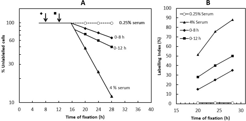
Attempts to map the Restriction Point in the mammalian cell cycle typically involve stimulating quiescent cells with mitogens for increasing intervals, removing the stimulus and then determining the proportion of cells that reach S phase at some point later. This "fixed point" estimate assumes that further cell cycle commitment ceases as soon as the stimulus is removed. In fact, kinetic analysis shows that the probability of cell cycle commitment does not fall back to its initial low value, immediately after a pulse of mitogens, but may instead remain slightly elevated for some while afterwards, compared to the starting quiescent population. Thus, cells entering S phase after a brief exposure to mitogens are not those that pass the Restriction Point early. Rather, they represent cells that continue on to S phase as a result of this residual, low probability of cell cycle commitment. Instead, the mitogen-regulated process(es) affecting the probability of cell cycle commitment are much closer to the start of S phase itself. Since the acquisition of (apparent) mitogen independence is such a poor indicator of the timing of cell cycle commitment, it is argued that a better measure is the point of insensitivity to CDK4,6 inhibitors such as palbociclib, which indicates when hyperphosphorylation of the Retinoblastoma Protein, RB, ceases to be dependent on mitogen-signalling pathways regulating CDK4,6/cyclin D activity.
期刊介绍:
Cell Division is an open access, peer-reviewed journal that encompasses all the molecular aspects of cell cycle control and cancer, cell growth, proliferation, survival, differentiation, signalling, gene transcription, protein synthesis, genome integrity, chromosome stability, centrosome duplication, DNA damage and DNA repair.
Cell Division provides an online forum for the cell-cycle community that aims to publish articles on all exciting aspects of cell-cycle research and to bridge the gap between models of cell cycle regulation, development, and cancer biology. This forum is driven by specialized and timely research articles, reviews and commentaries focused on this fast moving field, providing an invaluable tool for cell-cycle biologists.
Cell Division publishes articles in areas which includes, but not limited to:
DNA replication, cell fate decisions, cell cycle & development
Cell proliferation, mitosis, spindle assembly checkpoint, ubiquitin mediated degradation
DNA damage & repair
Apoptosis & cell death


 分享
分享
 求助内容:
求助内容: 应助结果提醒方式:
 扫码关注我们
