A detailed redescription of the mesoderm/neural crest cell boundary in the murine orbitotemporal region integrates the mammalian cranium into a pan-amniote cranial configuration
{"title":"A detailed redescription of the mesoderm/neural crest cell boundary in the murine orbitotemporal region integrates the mammalian cranium into a pan-amniote cranial configuration","authors":"Shunya Kuroda, Noritaka Adachi, Shigeru Kuratani","doi":"10.1111/ede.12411","DOIUrl":null,"url":null,"abstract":"<p>The morphology of the mammalian chondrocranium appears to differ significantly from those of other amniotes, since the former possesses uniquely developed brain and cranial sensory organs. In particular, a question has long remained unanswered as to the developmental and evolutionary origins of a cartilaginous nodule called the ala hypochiasmatica. In this study, we investigated the embryonic origin of skeletal elements in the murine orbitotemporal region by combining genetic cell lineage analysis with detailed morphological observation. Our results showed that the mesodermal embryonic environment including the ala hypochiasmatica, which appeared as an isolated mesodermal distribution in the neural crest-derived prechordal region, is formed as a part of the mesoderm that continued from the chordal region during early chondrocranial development. The mesoderm/neural crest cell boundary in the head mesenchyme is modified through development, resulting in the secondary mesodermal expansion to invade into the prechordal region. We thus revealed that the ala hypochiasmatica develops as the frontier of the mesodermal sheet stretched along the cephalic flexure. These results suggest that the mammalian ala hypochiasmatica has evolved from a part of the mesodermal primary cranial wall in ancestral amniotes. In addition, the endoskeletal elements in the orbitotemporal region, such as the orbital cartilage, suprapterygoid articulation of the palatoquadrate, and trabecula, some of which were once believed to represent primitive traits of amniotes and to be lost in the mammalian lineage, have been confirmed to exist in the mammalian cranium. Consequently, the mammalian chondrocranium can now be explained in relation to the pan-amniote cranial configuration.</p>","PeriodicalId":12083,"journal":{"name":"Evolution & Development","volume":"25 1","pages":"32-53"},"PeriodicalIF":2.6000,"publicationDate":"2022-07-31","publicationTypes":"Journal Article","fieldsOfStudy":null,"isOpenAccess":false,"openAccessPdf":"","citationCount":"2","resultStr":null,"platform":"Semanticscholar","paperid":null,"PeriodicalName":"Evolution & Development","FirstCategoryId":"99","ListUrlMain":"https://onlinelibrary.wiley.com/doi/10.1111/ede.12411","RegionNum":3,"RegionCategory":"生物学","ArticlePicture":[],"TitleCN":null,"AbstractTextCN":null,"PMCID":null,"EPubDate":"","PubModel":"","JCR":"Q2","JCRName":"DEVELOPMENTAL BIOLOGY","Score":null,"Total":0}
引用次数: 2
Abstract
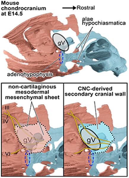
The morphology of the mammalian chondrocranium appears to differ significantly from those of other amniotes, since the former possesses uniquely developed brain and cranial sensory organs. In particular, a question has long remained unanswered as to the developmental and evolutionary origins of a cartilaginous nodule called the ala hypochiasmatica. In this study, we investigated the embryonic origin of skeletal elements in the murine orbitotemporal region by combining genetic cell lineage analysis with detailed morphological observation. Our results showed that the mesodermal embryonic environment including the ala hypochiasmatica, which appeared as an isolated mesodermal distribution in the neural crest-derived prechordal region, is formed as a part of the mesoderm that continued from the chordal region during early chondrocranial development. The mesoderm/neural crest cell boundary in the head mesenchyme is modified through development, resulting in the secondary mesodermal expansion to invade into the prechordal region. We thus revealed that the ala hypochiasmatica develops as the frontier of the mesodermal sheet stretched along the cephalic flexure. These results suggest that the mammalian ala hypochiasmatica has evolved from a part of the mesodermal primary cranial wall in ancestral amniotes. In addition, the endoskeletal elements in the orbitotemporal region, such as the orbital cartilage, suprapterygoid articulation of the palatoquadrate, and trabecula, some of which were once believed to represent primitive traits of amniotes and to be lost in the mammalian lineage, have been confirmed to exist in the mammalian cranium. Consequently, the mammalian chondrocranium can now be explained in relation to the pan-amniote cranial configuration.
期刊介绍:
Evolution & Development serves as a voice for the rapidly growing research community at the interface of evolutionary and developmental biology. The exciting re-integration of these two fields, after almost a century''s separation, holds much promise as the focus of a broader synthesis of biological thought. Evolution & Development publishes works that address the evolution/development interface from a diversity of angles. The journal welcomes papers from paleontologists, population biologists, developmental biologists, and molecular biologists, but also encourages submissions from professionals in other fields where relevant research is being carried out, from mathematics to the history and philosophy of science.


 分享
分享
 求助内容:
求助内容: 应助结果提醒方式:
 扫码关注我们
