{"title":"Primary Isolated Breast Lymphoma Presenting as Primary Breast Cancer with <sup>18</sup>F-FDG PET/CT.","authors":"Özge Vural Topuz, Özgür Omak, Burçak Yılmaz","doi":"10.4274/mirt.galenos.2022.58671","DOIUrl":null,"url":null,"abstract":"<p><p>A 40-year-old woman with a palpable mass lesion in her right breast suggested as breast cancer was admitted to <sup>18</sup>F-fluorodeoxyglucose (FDG) positron emission tomography/computed tomography (PET/CT) unit for the metabolic characterization of the lesion and for the staging of the disease. The patient had no fever and no evidence of weight loss or night sweats. <sup>18</sup>F-FDG PET/CT revealed an isolated solid mass lesion with increased <sup>18</sup>F-FDG uptake in the upper outer quadrant of the right breast and increased <sup>18</sup>F-FDG uptake in the lymph nodes of the right axilla suspected as primary breast cancer and its local lymph node metastasis. There was no other pathological <sup>18</sup>F-FDG uptake in the whole body. Excisional biopsy histopathology revealed diffuse large B-cell non-Hodgkin lymphoma.</p>","PeriodicalId":44681,"journal":{"name":"Molecular Imaging and Radionuclide Therapy","volume":"32 1","pages":"83-86"},"PeriodicalIF":1.1000,"publicationDate":"2023-02-23","publicationTypes":"Journal Article","fieldsOfStudy":null,"isOpenAccess":false,"openAccessPdf":"https://ftp.ncbi.nlm.nih.gov/pub/pmc/oa_pdf/c0/de/MIRT-32-83.PMC9950683.pdf","citationCount":"0","resultStr":null,"platform":"Semanticscholar","paperid":null,"PeriodicalName":"Molecular Imaging and Radionuclide Therapy","FirstCategoryId":"1085","ListUrlMain":"https://doi.org/10.4274/mirt.galenos.2022.58671","RegionNum":0,"RegionCategory":null,"ArticlePicture":[],"TitleCN":null,"AbstractTextCN":null,"PMCID":null,"EPubDate":"","PubModel":"","JCR":"Q4","JCRName":"RADIOLOGY, NUCLEAR MEDICINE & MEDICAL IMAGING","Score":null,"Total":0}
引用次数: 0
Abstract
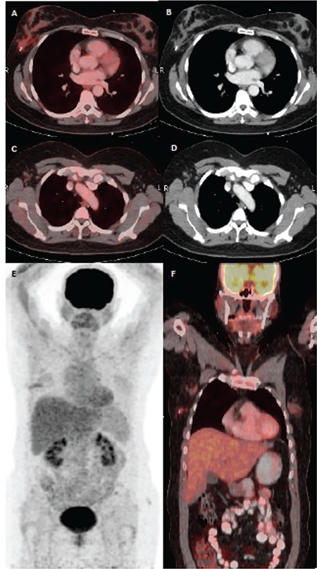
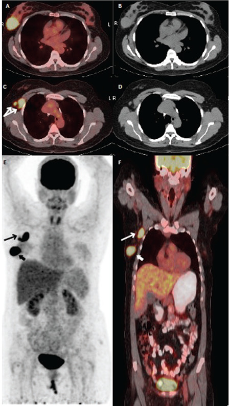
A 40-year-old woman with a palpable mass lesion in her right breast suggested as breast cancer was admitted to 18F-fluorodeoxyglucose (FDG) positron emission tomography/computed tomography (PET/CT) unit for the metabolic characterization of the lesion and for the staging of the disease. The patient had no fever and no evidence of weight loss or night sweats. 18F-FDG PET/CT revealed an isolated solid mass lesion with increased 18F-FDG uptake in the upper outer quadrant of the right breast and increased 18F-FDG uptake in the lymph nodes of the right axilla suspected as primary breast cancer and its local lymph node metastasis. There was no other pathological 18F-FDG uptake in the whole body. Excisional biopsy histopathology revealed diffuse large B-cell non-Hodgkin lymphoma.
期刊介绍:
Molecular Imaging and Radionuclide Therapy (Mol Imaging Radionucl Ther, MIRT) is publishes original research articles, invited reviews, editorials, short communications, letters, consensus statements, guidelines and case reports with a literature review on the topic, in the field of molecular imaging, multimodality imaging, nuclear medicine, radionuclide therapy, radiopharmacy, medical physics, dosimetry and radiobiology.


 分享
分享
 求助内容:
求助内容: 应助结果提醒方式:
 扫码关注我们
