{"title":"Correlation between gray values in cone-beam computed tomography and histomorphometric analysis.","authors":"Najmeh Anbiaee, Reihaneh Shafieian, Farid Shiezadeh, Mohammadtaghi Shakeri, Fatemeh Naqipour","doi":"10.5624/isd.20220051","DOIUrl":null,"url":null,"abstract":"<p><strong>Purpose: </strong>The aim of this study was to analyze the relationships between bone density measurements obtained using cone-beam computed tomography (CBCT) and morphometric parameters of bone determined by histomorphometric analysis.</p><p><strong>Materials and methods: </strong>In this <i>in vivo</i> study, 30 samples from the maxillary bones of 7 sheep were acquired using a trephine. The bone samples were returned to their original sites, and the sheep heads were imaged using CBCT. On the CBCT images, gray values were calculated. In the histomorphometric analysis, the total bone volume, the trabecular bone volume (referred to simply as bone volume), and the trabecular thickness were assessed.</p><p><strong>Results: </strong>Statistical testing showed significant correlations between CBCT gray values and total bone volume (r=0.537, <i>P</i>=0.002), bone volume (r=0.672, <i>P</i><0.001), and trabecular thickness (r=0.692, <i>P</i><0.001), as determined via the histomorphometric analysis.</p><p><strong>Conclusion: </strong>The results indicate a significant and acceptable association between CBCT gray values and bone volume, suggesting that CBCT may be used in bone densitometry.</p>","PeriodicalId":51714,"journal":{"name":"Imaging Science in Dentistry","volume":"52 4","pages":"375-382"},"PeriodicalIF":2.1000,"publicationDate":"2022-12-01","publicationTypes":"Journal Article","fieldsOfStudy":null,"isOpenAccess":false,"openAccessPdf":"https://ftp.ncbi.nlm.nih.gov/pub/pmc/oa_pdf/4f/4b/isd-52-375.PMC9807799.pdf","citationCount":"1","resultStr":null,"platform":"Semanticscholar","paperid":null,"PeriodicalName":"Imaging Science in Dentistry","FirstCategoryId":"1085","ListUrlMain":"https://doi.org/10.5624/isd.20220051","RegionNum":0,"RegionCategory":null,"ArticlePicture":[],"TitleCN":null,"AbstractTextCN":null,"PMCID":null,"EPubDate":"","PubModel":"","JCR":"Q3","JCRName":"DENTISTRY, ORAL SURGERY & MEDICINE","Score":null,"Total":0}
引用次数: 1
Abstract
Purpose: The aim of this study was to analyze the relationships between bone density measurements obtained using cone-beam computed tomography (CBCT) and morphometric parameters of bone determined by histomorphometric analysis.
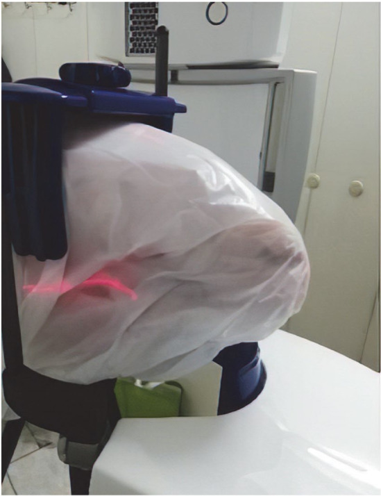
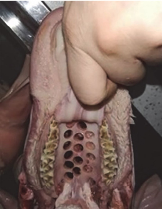
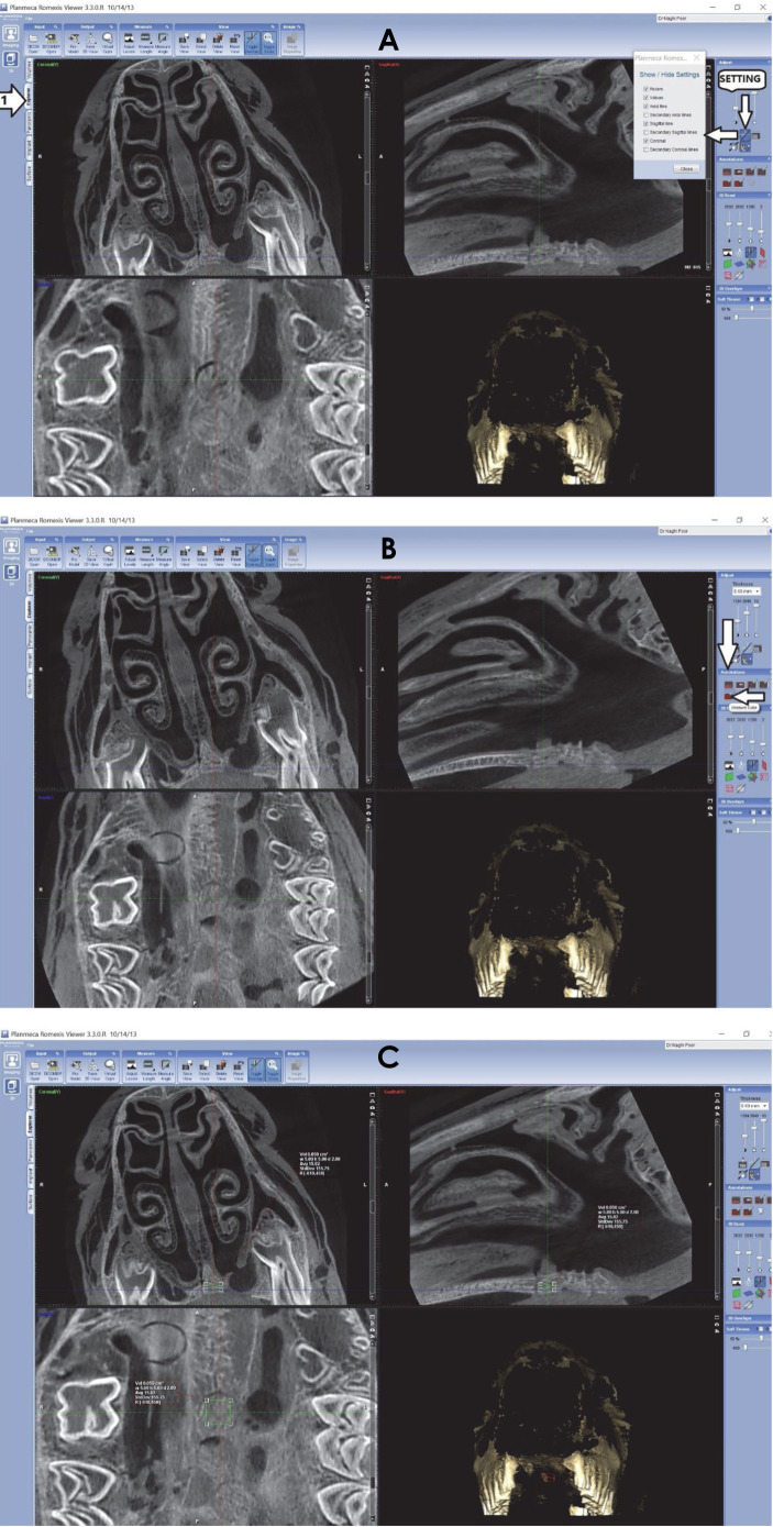
Materials and methods: In this in vivo study, 30 samples from the maxillary bones of 7 sheep were acquired using a trephine. The bone samples were returned to their original sites, and the sheep heads were imaged using CBCT. On the CBCT images, gray values were calculated. In the histomorphometric analysis, the total bone volume, the trabecular bone volume (referred to simply as bone volume), and the trabecular thickness were assessed.
Results: Statistical testing showed significant correlations between CBCT gray values and total bone volume (r=0.537, P=0.002), bone volume (r=0.672, P<0.001), and trabecular thickness (r=0.692, P<0.001), as determined via the histomorphometric analysis.
Conclusion: The results indicate a significant and acceptable association between CBCT gray values and bone volume, suggesting that CBCT may be used in bone densitometry.


 分享
分享
 求助内容:
求助内容: 应助结果提醒方式:
 扫码关注我们
