{"title":"Analysis of the root position and angulation of maxillary premolars in alveolar bone using cone-beam computed tomography.","authors":"Yun-Hoa Jung, Bong-Hae Cho, Jae-Joon Hwang","doi":"10.5624/isd.20220710","DOIUrl":null,"url":null,"abstract":"<p><strong>Purpose: </strong>This study investigated whether the relationship between the maxillary sinus and the root of the maxillary premolar is correlated with the root position and whether there is a difference in the long axis angle of premolars and the buccal bone thickness according to the sinus-root relationship and root position.</p><p><strong>Materials and methods: </strong>Cone-beam computed tomographic images of 587 maxillary first premolars and 580 second premolars from 303 patients were retrospectively reviewed. The maxillary sinus floor-root relationship was classified into 4 types, and the root position in the alveolar bone was evaluated as buccal, middle, or palatal. The long axis angle of the maxillary premolars in the alveolar bone and the buccal bone thickness were measured. The correlation between these parameters was analyzed.</p><p><strong>Results: </strong>The maxillary sinus floor-root relationship showed a statistically significant correlation with the root position in the alveolar bone. Most maxillary first premolars were buccally located, and more than half of the second premolars had their roots in the middle. The long axis angle of the premolars was significantly larger in buccal-positioned teeth than in middle-positioned teeth, and the buccal bone was thinner.</p><p><strong>Conclusion: </strong>When the root of the maxillary premolar was separated from the sinus floor, the premolar was often located on the buccal side. Most of the maxillary first premolars had a thinner buccal bone and larger inclination than the second premolars. It is recommended to evaluate the root position, sagittal angle and buccal bone thickness using CBCT for implant treatment planning.</p>","PeriodicalId":51714,"journal":{"name":"Imaging Science in Dentistry","volume":"52 4","pages":"365-373"},"PeriodicalIF":2.1000,"publicationDate":"2022-12-01","publicationTypes":"Journal Article","fieldsOfStudy":null,"isOpenAccess":false,"openAccessPdf":"https://ftp.ncbi.nlm.nih.gov/pub/pmc/oa_pdf/13/9d/isd-52-365.PMC9807791.pdf","citationCount":"0","resultStr":null,"platform":"Semanticscholar","paperid":null,"PeriodicalName":"Imaging Science in Dentistry","FirstCategoryId":"1085","ListUrlMain":"https://doi.org/10.5624/isd.20220710","RegionNum":0,"RegionCategory":null,"ArticlePicture":[],"TitleCN":null,"AbstractTextCN":null,"PMCID":null,"EPubDate":"","PubModel":"","JCR":"Q3","JCRName":"DENTISTRY, ORAL SURGERY & MEDICINE","Score":null,"Total":0}
引用次数: 0
Abstract
Purpose: This study investigated whether the relationship between the maxillary sinus and the root of the maxillary premolar is correlated with the root position and whether there is a difference in the long axis angle of premolars and the buccal bone thickness according to the sinus-root relationship and root position.
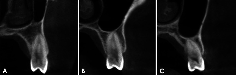
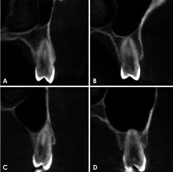
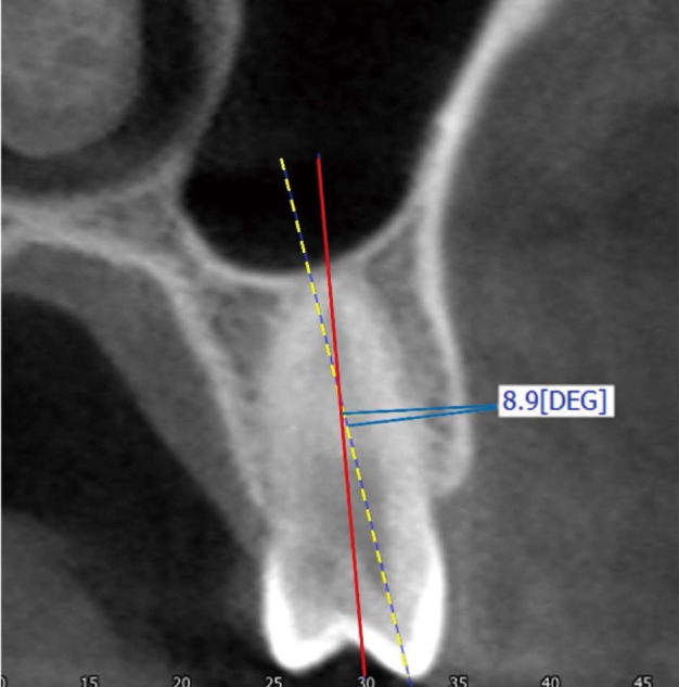
Materials and methods: Cone-beam computed tomographic images of 587 maxillary first premolars and 580 second premolars from 303 patients were retrospectively reviewed. The maxillary sinus floor-root relationship was classified into 4 types, and the root position in the alveolar bone was evaluated as buccal, middle, or palatal. The long axis angle of the maxillary premolars in the alveolar bone and the buccal bone thickness were measured. The correlation between these parameters was analyzed.
Results: The maxillary sinus floor-root relationship showed a statistically significant correlation with the root position in the alveolar bone. Most maxillary first premolars were buccally located, and more than half of the second premolars had their roots in the middle. The long axis angle of the premolars was significantly larger in buccal-positioned teeth than in middle-positioned teeth, and the buccal bone was thinner.
Conclusion: When the root of the maxillary premolar was separated from the sinus floor, the premolar was often located on the buccal side. Most of the maxillary first premolars had a thinner buccal bone and larger inclination than the second premolars. It is recommended to evaluate the root position, sagittal angle and buccal bone thickness using CBCT for implant treatment planning.


 分享
分享
 求助内容:
求助内容: 应助结果提醒方式:
 扫码关注我们
