Emel Tuğba Ataman-Duruel, Zehra Beycioğlu, Doğukan Yılmaz, Samir Goyushov, Tansu Çimen, Onurcem Duruel, Hasan Güney Yılmaz, Tolga Fikret Tözüm
{"title":"Evaluation of Cortical Thicknesses and Bone Density Values of Mandibular Canal Borders and Coronal Site of Alveolar Crest.","authors":"Emel Tuğba Ataman-Duruel, Zehra Beycioğlu, Doğukan Yılmaz, Samir Goyushov, Tansu Çimen, Onurcem Duruel, Hasan Güney Yılmaz, Tolga Fikret Tözüm","doi":"10.5037/jomr.2023.14304","DOIUrl":null,"url":null,"abstract":"<p><strong>Objectives: </strong>The objectives of this retrospective study are to measure the amount of the alveolar crest cortication and cortication around the mandibular canal, and to evaluate bone density values of alveolar crest, cortication around mandibular canal, and possible implant placement area for edentulous sites.</p><p><strong>Material and methods: </strong>Six hundred forty-two cone-beam computed tomography scans from 642 subjects were evaluated in four centers. Cortical thicknesses of alveolar crest and mandibular canal cortical borders (buccal, lingual, apical, and coronal) in each mandibular posterior teeth region were measured. Bone density of alveolar crest and mandibular canal cortical borders (buccal, lingual, apical, and coronal) in each mandibular posterior teeth region were recorded. The correlations between numeric variables were investigated using Pearson's correlation test.</p><p><strong>Results: </strong>The largest cortical border of the canal was measured 1.1 (SD 0.71) mm at the left second molar area and in coronal side of the mandibular canal (MC). Left and right first premolar regions showed higher bone density values compared to the other sites in all bone density values evaluations. The buccal side of the canal at the right first premolar region showed the highest bone density values (832.32 [SD 350.01]) while the coronal side of the canal at the left second molar region showed the lowest (508.75 [SD 225.47]). The bone density of possible implant placement area at the both left (692.25 [SD 238.25]) and right (604.43 [SD 240.92]) edentulous first premolar showed the highest values. Positive correlations between the bone density values of alveolar crest and the coronal side of MC were found in molar and left second premolar regions (P < 0.05).</p><p><strong>Conclusions: </strong>Results may provide information about the amount of cortication and bone densities tooth by tooth for posterior mandible to surgeons for planning the treatment precisely.</p>","PeriodicalId":53254,"journal":{"name":"eJournal of Oral Maxillofacial Research","volume":"14 3","pages":"e4"},"PeriodicalIF":1.0000,"publicationDate":"2023-09-30","publicationTypes":"Journal Article","fieldsOfStudy":null,"isOpenAccess":false,"openAccessPdf":"https://www.ncbi.nlm.nih.gov/pmc/articles/PMC10645474/pdf/","citationCount":"0","resultStr":null,"platform":"Semanticscholar","paperid":null,"PeriodicalName":"eJournal of Oral Maxillofacial Research","FirstCategoryId":"1085","ListUrlMain":"https://doi.org/10.5037/jomr.2023.14304","RegionNum":0,"RegionCategory":null,"ArticlePicture":[],"TitleCN":null,"AbstractTextCN":null,"PMCID":null,"EPubDate":"2023/7/1 0:00:00","PubModel":"eCollection","JCR":"Q3","JCRName":"DENTISTRY, ORAL SURGERY & MEDICINE","Score":null,"Total":0}
引用次数: 0
Abstract
Objectives: The objectives of this retrospective study are to measure the amount of the alveolar crest cortication and cortication around the mandibular canal, and to evaluate bone density values of alveolar crest, cortication around mandibular canal, and possible implant placement area for edentulous sites.
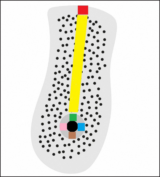
Material and methods: Six hundred forty-two cone-beam computed tomography scans from 642 subjects were evaluated in four centers. Cortical thicknesses of alveolar crest and mandibular canal cortical borders (buccal, lingual, apical, and coronal) in each mandibular posterior teeth region were measured. Bone density of alveolar crest and mandibular canal cortical borders (buccal, lingual, apical, and coronal) in each mandibular posterior teeth region were recorded. The correlations between numeric variables were investigated using Pearson's correlation test.
Results: The largest cortical border of the canal was measured 1.1 (SD 0.71) mm at the left second molar area and in coronal side of the mandibular canal (MC). Left and right first premolar regions showed higher bone density values compared to the other sites in all bone density values evaluations. The buccal side of the canal at the right first premolar region showed the highest bone density values (832.32 [SD 350.01]) while the coronal side of the canal at the left second molar region showed the lowest (508.75 [SD 225.47]). The bone density of possible implant placement area at the both left (692.25 [SD 238.25]) and right (604.43 [SD 240.92]) edentulous first premolar showed the highest values. Positive correlations between the bone density values of alveolar crest and the coronal side of MC were found in molar and left second premolar regions (P < 0.05).
Conclusions: Results may provide information about the amount of cortication and bone densities tooth by tooth for posterior mandible to surgeons for planning the treatment precisely.


 分享
分享

 求助内容:
求助内容: 应助结果提醒方式:
 扫码关注我们
