{"title":"Issue Information","authors":"","doi":"10.1002/cpsc.91","DOIUrl":null,"url":null,"abstract":"<p><b>Cover</b>: In Döpper et al. (http://doi.org/10.1002/cpsc.120), the image shows immunostaining for early retinal markers and all retinal cell types. At d35, the outer layer stains positive for the early eye field marker RX. Moreover, in the outer part, retinal progenitor cells (VSX2), and in the inner part, ganglion cells are present (BRN3). CRX, a marker for retinal progenitors and immature photoreceptors, is present at d35, and a shift to the outer layer can be detected at d61. Horizontal cells (PROX1) and amacrine cells (AP2α) are present in the inner nuclear layer (d61, d126). Cone photoreceptors (RXRγ, ARR3; d96, d126, d152) and rod photoreceptors (NRL; d152) are present in the outer nuclear layer. Bipolar cells (PKCα) and Müller glia cells (VIM) are present in the inner nuclear layer (d152). A schematic overview of the retina with the seven different cell types organized in specific layers is given. Secondary antibodies were labeled in green (AlexaFluor 488), and nuclei were stained with DAPI (blue). Scale bar: 50 µm.\n\n <figure>\n <div><picture>\n <source></source></picture><p></p>\n </div>\n </figure></p>","PeriodicalId":53703,"journal":{"name":"Current Protocols in Stem Cell Biology","volume":"55 1","pages":""},"PeriodicalIF":0.0000,"publicationDate":"2020-09-21","publicationTypes":"Journal Article","fieldsOfStudy":null,"isOpenAccess":false,"openAccessPdf":"https://sci-hub-pdf.com/10.1002/cpsc.91","citationCount":"0","resultStr":null,"platform":"Semanticscholar","paperid":null,"PeriodicalName":"Current Protocols in Stem Cell Biology","FirstCategoryId":"1085","ListUrlMain":"https://onlinelibrary.wiley.com/doi/10.1002/cpsc.91","RegionNum":0,"RegionCategory":null,"ArticlePicture":[],"TitleCN":null,"AbstractTextCN":null,"PMCID":null,"EPubDate":"","PubModel":"","JCR":"Q2","JCRName":"Biochemistry, Genetics and Molecular Biology","Score":null,"Total":0}
引用次数: 0
Abstract
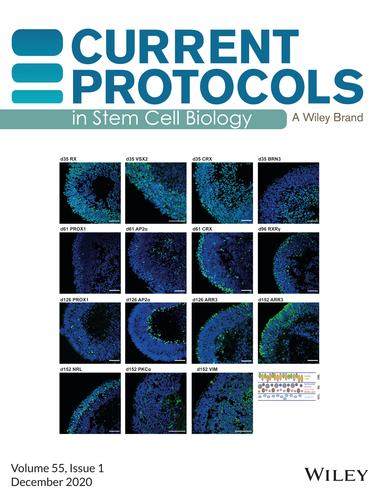
Cover: In Döpper et al. (http://doi.org/10.1002/cpsc.120), the image shows immunostaining for early retinal markers and all retinal cell types. At d35, the outer layer stains positive for the early eye field marker RX. Moreover, in the outer part, retinal progenitor cells (VSX2), and in the inner part, ganglion cells are present (BRN3). CRX, a marker for retinal progenitors and immature photoreceptors, is present at d35, and a shift to the outer layer can be detected at d61. Horizontal cells (PROX1) and amacrine cells (AP2α) are present in the inner nuclear layer (d61, d126). Cone photoreceptors (RXRγ, ARR3; d96, d126, d152) and rod photoreceptors (NRL; d152) are present in the outer nuclear layer. Bipolar cells (PKCα) and Müller glia cells (VIM) are present in the inner nuclear layer (d152). A schematic overview of the retina with the seven different cell types organized in specific layers is given. Secondary antibodies were labeled in green (AlexaFluor 488), and nuclei were stained with DAPI (blue). Scale bar: 50 µm.
期刊介绍:
Published in affiliation with the International Society for Stem Cell Research (ISSCR), Current Protocols in Stem Cell Biology (CPSC) covers the most fundamental protocols and methods in the rapidly growing field of stem cell biology. Updated monthly, CPSC will constantly evolve with thelatest developments and breakthroughs in the field. Drawing on the expertise of leading researchers from around the world, Current Protocols in Stem Cell Biology includes methods and insights that will enhance the progress of global research.


 分享
分享
 求助内容:
求助内容: 应助结果提醒方式:
 扫码关注我们
