Nikolai P. Melnikov, Fyodor V. Bolshakov, Veronika S. Frolova, Kseniia V. Skorentseva, Alexander V. Ereskovsky, Alina A. Saidova, Andrey I. Lavrov
{"title":"Tissue homeostasis in sponges: Quantitative analysis of cell proliferation and apoptosis","authors":"Nikolai P. Melnikov, Fyodor V. Bolshakov, Veronika S. Frolova, Kseniia V. Skorentseva, Alexander V. Ereskovsky, Alina A. Saidova, Andrey I. Lavrov","doi":"10.1002/jez.b.23138","DOIUrl":null,"url":null,"abstract":"<p>Tissues of multicellular animals are maintained due to a tight balance between cell proliferation and programmed cell death. Sponges are early branching metazoans essential to understanding the key mechanisms of tissue homeostasis. This article is dedicated to the comparative analysis of proliferation and apoptosis in intact tissues of two sponges, <i>Halisarca dujardinii</i> (class Demospongiae) and <i>Leucosolenia variabilis</i> (class Calcarea). Labeled nucleotides EdU and anti-phosphorylated histone 3 antibodies reveal a considerable number of cycling cells in intact tissues of both species. Quantitative DNA staining reveals the classic cell cycle distribution curve. The main type of cycling cells are choanocytes - flagellated cells of the aquiferous system. The rate of proliferation remains constant throughout various areas of sponge bodies that contain choanocytes. The EdU tracking experiments conducted in <i>H. dujardinii</i> indicate that choanocytes may give rise to mesohyl cells through migration. The number of apoptotic cells in tissues of both species is insignificant, although being comparable to the renewing tissues of other animals. <i>In vivo</i> studies with tetramethylrhodamine ethyl ester and CellEvent Caspase-3/7 indicate that apoptosis might be independent of mitochondrial outer membrane permeabilization. Altogether, a combination of confocal laser scanning microscopy and flow cytometry provides a quantitative description of cell proliferation and apoptosis in sponges displaying either rapid growth or cell turnover.</p>","PeriodicalId":15682,"journal":{"name":"Journal of experimental zoology. Part B, Molecular and developmental evolution","volume":"338 6","pages":"360-381"},"PeriodicalIF":1.7000,"publicationDate":"2022-04-25","publicationTypes":"Journal Article","fieldsOfStudy":null,"isOpenAccess":false,"openAccessPdf":"","citationCount":"4","resultStr":null,"platform":"Semanticscholar","paperid":null,"PeriodicalName":"Journal of experimental zoology. Part B, Molecular and developmental evolution","FirstCategoryId":"99","ListUrlMain":"https://onlinelibrary.wiley.com/doi/10.1002/jez.b.23138","RegionNum":3,"RegionCategory":"生物学","ArticlePicture":[],"TitleCN":null,"AbstractTextCN":null,"PMCID":null,"EPubDate":"","PubModel":"","JCR":"Q3","JCRName":"DEVELOPMENTAL BIOLOGY","Score":null,"Total":0}
引用次数: 4
Abstract
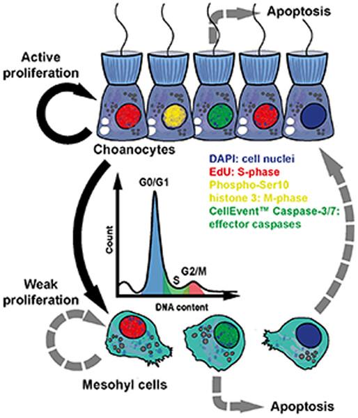
Tissues of multicellular animals are maintained due to a tight balance between cell proliferation and programmed cell death. Sponges are early branching metazoans essential to understanding the key mechanisms of tissue homeostasis. This article is dedicated to the comparative analysis of proliferation and apoptosis in intact tissues of two sponges, Halisarca dujardinii (class Demospongiae) and Leucosolenia variabilis (class Calcarea). Labeled nucleotides EdU and anti-phosphorylated histone 3 antibodies reveal a considerable number of cycling cells in intact tissues of both species. Quantitative DNA staining reveals the classic cell cycle distribution curve. The main type of cycling cells are choanocytes - flagellated cells of the aquiferous system. The rate of proliferation remains constant throughout various areas of sponge bodies that contain choanocytes. The EdU tracking experiments conducted in H. dujardinii indicate that choanocytes may give rise to mesohyl cells through migration. The number of apoptotic cells in tissues of both species is insignificant, although being comparable to the renewing tissues of other animals. In vivo studies with tetramethylrhodamine ethyl ester and CellEvent Caspase-3/7 indicate that apoptosis might be independent of mitochondrial outer membrane permeabilization. Altogether, a combination of confocal laser scanning microscopy and flow cytometry provides a quantitative description of cell proliferation and apoptosis in sponges displaying either rapid growth or cell turnover.
期刊介绍:
Developmental Evolution is a branch of evolutionary biology that integrates evidence and concepts from developmental biology, phylogenetics, comparative morphology, evolutionary genetics and increasingly also genomics, systems biology as well as synthetic biology to gain an understanding of the structure and evolution of organisms.
The Journal of Experimental Zoology -B: Molecular and Developmental Evolution provides a forum where these fields are invited to bring together their insights to further a synthetic understanding of evolution from the molecular through the organismic level. Contributions from all these branches of science are welcome to JEZB.
We particularly encourage submissions that apply the tools of genomics, as well as systems and synthetic biology to developmental evolution. At this time the impact of these emerging fields on developmental evolution has not been explored to its fullest extent and for this reason we are eager to foster the relationship of systems and synthetic biology with devo evo.


 分享
分享
 求助内容:
求助内容: 应助结果提醒方式:
 扫码关注我们
