{"title":"Motion Characterization of Pacemaker Lead Wire In Vivo for Piezoelectric Energy Harvesting Applications.","authors":"Christopher Hu, Kamran Behdinan","doi":"10.1007/s13239-023-00700-3","DOIUrl":null,"url":null,"abstract":"<p><strong>Purpose: </strong>Piezoelectric energy harvesters (PEH) for cardiac pacemakers typically use animal models to assess the performance of the PEH. However, if considering multiple designs, the use of animal models and prototyping increases costs and time. To reduce the use of animal models in research for pacemaker energy harvesting applications, this study investigates the motion of a pacemaker lead wire (PLW) in vivo using fluoroscopy imaging to quantify the position and displacements as a function of time, such that the data can be used in computer simulations.</p><p><strong>Methods: </strong>The proposed technique uses fluoroscopy imaging video data of a dual chamber pacemaker implanted in a patient, and image processing allows for the motion of the PLW captured. The motion is discretized into nodes for ease of implementation in finite element software. FEA simulation is presented using a piezoelectric energy harvester design integrated in the lead wire, and the energy output is predicted by finite element computer simulation.</p><p><strong>Results: </strong>A 2-dimensional analysis is conducted with the fluoroscopy imaging video data to characterize the PLW motion and results show close agreement with literature values. Simulations with an energy harvesting circuit using the nodal position and displacement data shows that a PEH integrated in the PLW can generate a direct current voltage of 1.12 V and power output of 0.125 μW, potentially extending the battery life of pacemakers by 0.75-1 years.</p><p><strong>Conclusions: </strong>The results suggest that fluoroscopy imaging data can be effective in evaluating PEH designs rather than using animal models, saving time and costs.</p>","PeriodicalId":54322,"journal":{"name":"Cardiovascular Engineering and Technology","volume":" ","pages":"111-122"},"PeriodicalIF":1.8000,"publicationDate":"2024-04-01","publicationTypes":"Journal Article","fieldsOfStudy":null,"isOpenAccess":false,"openAccessPdf":"","citationCount":"0","resultStr":null,"platform":"Semanticscholar","paperid":null,"PeriodicalName":"Cardiovascular Engineering and Technology","FirstCategoryId":"5","ListUrlMain":"https://doi.org/10.1007/s13239-023-00700-3","RegionNum":4,"RegionCategory":"医学","ArticlePicture":[],"TitleCN":null,"AbstractTextCN":null,"PMCID":null,"EPubDate":"2023/11/22 0:00:00","PubModel":"Epub","JCR":"Q3","JCRName":"CARDIAC & CARDIOVASCULAR SYSTEMS","Score":null,"Total":0}
引用次数: 0
Abstract
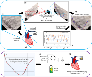
Purpose: Piezoelectric energy harvesters (PEH) for cardiac pacemakers typically use animal models to assess the performance of the PEH. However, if considering multiple designs, the use of animal models and prototyping increases costs and time. To reduce the use of animal models in research for pacemaker energy harvesting applications, this study investigates the motion of a pacemaker lead wire (PLW) in vivo using fluoroscopy imaging to quantify the position and displacements as a function of time, such that the data can be used in computer simulations.
Methods: The proposed technique uses fluoroscopy imaging video data of a dual chamber pacemaker implanted in a patient, and image processing allows for the motion of the PLW captured. The motion is discretized into nodes for ease of implementation in finite element software. FEA simulation is presented using a piezoelectric energy harvester design integrated in the lead wire, and the energy output is predicted by finite element computer simulation.
Results: A 2-dimensional analysis is conducted with the fluoroscopy imaging video data to characterize the PLW motion and results show close agreement with literature values. Simulations with an energy harvesting circuit using the nodal position and displacement data shows that a PEH integrated in the PLW can generate a direct current voltage of 1.12 V and power output of 0.125 μW, potentially extending the battery life of pacemakers by 0.75-1 years.
Conclusions: The results suggest that fluoroscopy imaging data can be effective in evaluating PEH designs rather than using animal models, saving time and costs.
期刊介绍:
Cardiovascular Engineering and Technology is a journal publishing the spectrum of basic to translational research in all aspects of cardiovascular physiology and medical treatment. It is the forum for academic and industrial investigators to disseminate research that utilizes engineering principles and methods to advance fundamental knowledge and technological solutions related to the cardiovascular system. Manuscripts spanning from subcellular to systems level topics are invited, including but not limited to implantable medical devices, hemodynamics and tissue biomechanics, functional imaging, surgical devices, electrophysiology, tissue engineering and regenerative medicine, diagnostic instruments, transport and delivery of biologics, and sensors. In addition to manuscripts describing the original publication of research, manuscripts reviewing developments in these topics or their state-of-art are also invited.


 分享
分享
 求助内容:
求助内容: 应助结果提醒方式:
 扫码关注我们
