Sajad Sarvari, Duncan McGee, Ryan O'Connell, Oxana Tseytlin, Andrey A Bobko, Mark Tseytlin
{"title":"Electron Spin Resonance Probe Incorporation into Bioinks Permits Longitudinal Oxygen Imaging of Bioprinted Constructs.","authors":"Sajad Sarvari, Duncan McGee, Ryan O'Connell, Oxana Tseytlin, Andrey A Bobko, Mark Tseytlin","doi":"10.1007/s11307-023-01871-0","DOIUrl":null,"url":null,"abstract":"<p><strong>Purpose: </strong>Bioprinting is an additive manufacturing technology analogous to 3D printing. Instead of plastic or resin, cell-laden hydrogels are used to produce a construct of the intended biological structure. Over time, cells transform this construct into a functioning tissue or organ. The process of printing followed by tissue maturation is referred to as 4D bioprinting. The fourth dimension is temporal. Failure to provide living cells with sufficient amounts of oxygen at any point along the developmental timeline may jeopardize the bioprinting goals. Even transient hypoxia may alter cells' differentiation and proliferation or trigger apoptosis. Electron paramagnetic resonance (EPR) imaging modality is proposed to permit 4D monitoring of oxygen within bioprinted structures.</p><p><strong>Procedures: </strong>Lithium octa-n-butoxy-phthalocyanine (LiNc-BuO) probes have been introduced into gelatin methacrylate (GelMA) bioink. GelMA is a cross-linkable hydrogel, and LiNc-BuO is an oxygen-sensitive compound that permits longitudinal oximetric measurements. The effects of the oxygen probe on printability have been evaluated. A digital light processing (DLP) bioprinter was built in the laboratory. Bioprinting protocols have been developed that consider the optical properties of the GelMA/LiNc-BuO composites. Acellular and cell-laden constructs have been printed and imaged. The post-printing effect of residual photoinitiator on oxygen depletion has been investigated.</p><p><strong>Results: </strong>Models have been successfully printed using a lab-built bioprinter. Rapid scan EPR images reflective of the expected oxygen concentration levels have been acquired. An unreported problem of oxygen depletion in bioprinted constructs by the residual photoinitiator has been documented. EPR imaging is proposed as a control method for its removal. The oxygen consumption rates by HEK293T cells within a bioprinted cylinder have been imaged and quantified.</p><p><strong>Conclusions: </strong>The feasibility of the cointegration of 4D EPR imaging and 4D bioprinting has been demonstrated. The proof-of-concept experiments, which were conducted using oxygen probes loaded into GelMA, lay the foundation for a broad range of applications, such as bioprinting with many types of bioinks loaded with diverse varieties of molecular spin probes.</p>","PeriodicalId":18760,"journal":{"name":"Molecular Imaging and Biology","volume":" ","pages":"511-524"},"PeriodicalIF":2.5000,"publicationDate":"2024-06-01","publicationTypes":"Journal Article","fieldsOfStudy":null,"isOpenAccess":false,"openAccessPdf":"https://www.ncbi.nlm.nih.gov/pmc/articles/PMC11211156/pdf/","citationCount":"0","resultStr":null,"platform":"Semanticscholar","paperid":null,"PeriodicalName":"Molecular Imaging and Biology","FirstCategoryId":"3","ListUrlMain":"https://doi.org/10.1007/s11307-023-01871-0","RegionNum":4,"RegionCategory":"医学","ArticlePicture":[],"TitleCN":null,"AbstractTextCN":null,"PMCID":null,"EPubDate":"2023/12/1 0:00:00","PubModel":"Epub","JCR":"Q2","JCRName":"RADIOLOGY, NUCLEAR MEDICINE & MEDICAL IMAGING","Score":null,"Total":0}
引用次数: 0
Abstract
Purpose: Bioprinting is an additive manufacturing technology analogous to 3D printing. Instead of plastic or resin, cell-laden hydrogels are used to produce a construct of the intended biological structure. Over time, cells transform this construct into a functioning tissue or organ. The process of printing followed by tissue maturation is referred to as 4D bioprinting. The fourth dimension is temporal. Failure to provide living cells with sufficient amounts of oxygen at any point along the developmental timeline may jeopardize the bioprinting goals. Even transient hypoxia may alter cells' differentiation and proliferation or trigger apoptosis. Electron paramagnetic resonance (EPR) imaging modality is proposed to permit 4D monitoring of oxygen within bioprinted structures.
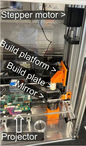
Procedures: Lithium octa-n-butoxy-phthalocyanine (LiNc-BuO) probes have been introduced into gelatin methacrylate (GelMA) bioink. GelMA is a cross-linkable hydrogel, and LiNc-BuO is an oxygen-sensitive compound that permits longitudinal oximetric measurements. The effects of the oxygen probe on printability have been evaluated. A digital light processing (DLP) bioprinter was built in the laboratory. Bioprinting protocols have been developed that consider the optical properties of the GelMA/LiNc-BuO composites. Acellular and cell-laden constructs have been printed and imaged. The post-printing effect of residual photoinitiator on oxygen depletion has been investigated.
Results: Models have been successfully printed using a lab-built bioprinter. Rapid scan EPR images reflective of the expected oxygen concentration levels have been acquired. An unreported problem of oxygen depletion in bioprinted constructs by the residual photoinitiator has been documented. EPR imaging is proposed as a control method for its removal. The oxygen consumption rates by HEK293T cells within a bioprinted cylinder have been imaged and quantified.
Conclusions: The feasibility of the cointegration of 4D EPR imaging and 4D bioprinting has been demonstrated. The proof-of-concept experiments, which were conducted using oxygen probes loaded into GelMA, lay the foundation for a broad range of applications, such as bioprinting with many types of bioinks loaded with diverse varieties of molecular spin probes.
期刊介绍:
Molecular Imaging and Biology (MIB) invites original contributions (research articles, review articles, commentaries, etc.) on the utilization of molecular imaging (i.e., nuclear imaging, optical imaging, autoradiography and pathology, MRI, MPI, ultrasound imaging, radiomics/genomics etc.) to investigate questions related to biology and health. The objective of MIB is to provide a forum to the discovery of molecular mechanisms of disease through the use of imaging techniques. We aim to investigate the biological nature of disease in patients and establish new molecular imaging diagnostic and therapy procedures.
Some areas that are covered are:
Preclinical and clinical imaging of macromolecular targets (e.g., genes, receptors, enzymes) involved in significant biological processes.
The design, characterization, and study of new molecular imaging probes and contrast agents for the functional interrogation of macromolecular targets.
Development and evaluation of imaging systems including instrumentation, image reconstruction algorithms, image analysis, and display.
Development of molecular assay approaches leading to quantification of the biological information obtained in molecular imaging.
Study of in vivo animal models of disease for the development of new molecular diagnostics and therapeutics.
Extension of in vitro and in vivo discoveries using disease models, into well designed clinical research investigations.
Clinical molecular imaging involving clinical investigations, clinical trials and medical management or cost-effectiveness studies.


 分享
分享

 求助内容:
求助内容: 应助结果提醒方式:
 扫码关注我们
