Thaiza Goncalves Rocha, Carla Barros de Oliveira, Roberto José Pessoa de Magalhães Filho, Angelo Maiolino, Marcela Baraúna Magno, Davi da Silva Barbirato, Eduardo Murad Villoria, Lucianne Cople Maia, Sandra Regina Torres, Maria Augusta Visconti
{"title":"Comparison between computed tomography and magnetic resonance imaging in detecting multiple myeloma lesions in the skull: A systematic review","authors":"Thaiza Goncalves Rocha, Carla Barros de Oliveira, Roberto José Pessoa de Magalhães Filho, Angelo Maiolino, Marcela Baraúna Magno, Davi da Silva Barbirato, Eduardo Murad Villoria, Lucianne Cople Maia, Sandra Regina Torres, Maria Augusta Visconti","doi":"10.1007/s40336-023-00605-0","DOIUrl":null,"url":null,"abstract":"<h3 data-test=\"abstract-sub-heading\">Objective</h3><p>Review, qualify and synthesize the evidence that compared computed tomography (CT) images with magnetic resonance imaging (MRI) in detecting multiple myeloma (MM) lesions in the skull, through a systematic review.</p><h3 data-test=\"abstract-sub-heading\">Methods</h3><p>Searches were performed in six databases and the grey literature, up to August 2023, without restriction by date or publication language. Observational studies comparing CT images and MRI of the skull of patients previously diagnosed with MM were included. Data were extracted by two reviewers in a standardized and independent manner. The methodological quality assessment was performed using the QUADAS-2 tool and the evidence certainty assessment using the GRADE tool.</p><h3 data-test=\"abstract-sub-heading\">Results</h3><p>Of the 911 identified references, 11 were included, and they all used either positron emission computed tomography (PET/CT) and/or low-dose computed tomography (LDCT) to compare to MRI. In 6 of 7 studies, MRI demonstrated a greater capacity to detect MM lesions than PET/CT images. When compared with LDCT images, MRI showed lower detection capacity in 4 studies. Six of the 11 included articles had a low risk of bias. However, as observational data evidence, the assessed certainty of the evidence was considered very low.</p><h3 data-test=\"abstract-sub-heading\">Conclusions</h3><p>PET/CT and MRI images presented limitations in detecting MM lesions in the skull compared to LDCT images. The evidence suggested that the greatest detection capability could be achieved by employing whole-body MRI complemented by LDCT images of the skull. Future studies are needed to confirm this result.</p>","PeriodicalId":48600,"journal":{"name":"Clinical and Translational Imaging","volume":"62 1","pages":""},"PeriodicalIF":1.6000,"publicationDate":"2023-12-07","publicationTypes":"Journal Article","fieldsOfStudy":null,"isOpenAccess":false,"openAccessPdf":"","citationCount":"0","resultStr":null,"platform":"Semanticscholar","paperid":null,"PeriodicalName":"Clinical and Translational Imaging","FirstCategoryId":"3","ListUrlMain":"https://doi.org/10.1007/s40336-023-00605-0","RegionNum":4,"RegionCategory":"医学","ArticlePicture":[],"TitleCN":null,"AbstractTextCN":null,"PMCID":null,"EPubDate":"","PubModel":"","JCR":"Q2","JCRName":"RADIOLOGY, NUCLEAR MEDICINE & MEDICAL IMAGING","Score":null,"Total":0}
引用次数: 0
Abstract
Objective
Review, qualify and synthesize the evidence that compared computed tomography (CT) images with magnetic resonance imaging (MRI) in detecting multiple myeloma (MM) lesions in the skull, through a systematic review.
Methods
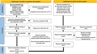
Searches were performed in six databases and the grey literature, up to August 2023, without restriction by date or publication language. Observational studies comparing CT images and MRI of the skull of patients previously diagnosed with MM were included. Data were extracted by two reviewers in a standardized and independent manner. The methodological quality assessment was performed using the QUADAS-2 tool and the evidence certainty assessment using the GRADE tool.
Results
Of the 911 identified references, 11 were included, and they all used either positron emission computed tomography (PET/CT) and/or low-dose computed tomography (LDCT) to compare to MRI. In 6 of 7 studies, MRI demonstrated a greater capacity to detect MM lesions than PET/CT images. When compared with LDCT images, MRI showed lower detection capacity in 4 studies. Six of the 11 included articles had a low risk of bias. However, as observational data evidence, the assessed certainty of the evidence was considered very low.
Conclusions
PET/CT and MRI images presented limitations in detecting MM lesions in the skull compared to LDCT images. The evidence suggested that the greatest detection capability could be achieved by employing whole-body MRI complemented by LDCT images of the skull. Future studies are needed to confirm this result.
期刊介绍:
Clinical and Translational Imaging is an international journal that publishes timely, up-to-date summaries on clinical practice and translational research and clinical applications of approved and experimental radiopharmaceuticals for diagnostic and therapeutic purposes. Coverage includes such topics as advanced preclinical evidence in the fields of physics, dosimetry, radiation biology and radiopharmacy with relevance to applications in human subjects. The journal benefits a readership of nuclear medicine practitioners and allied professionals involved in molecular imaging and therapy.


 分享
分享
 求助内容:
求助内容: 应助结果提醒方式:
 扫码关注我们
