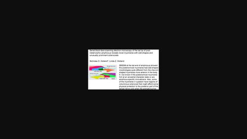
Serial block-face scanning electron microscopy of the tail tip of post-metamorphic amphioxus finds novel myomeres with odd shapes and unusually prominent sclerocoels
{"title":"Serial block-face scanning electron microscopy of the tail tip of post-metamorphic amphioxus finds novel myomeres with odd shapes and unusually prominent sclerocoels","authors":"Nicholas D. Holland, Linda Z. Holland","doi":"10.1002/jmor.21667","DOIUrl":null,"url":null,"abstract":"<p>Serial block-face scanning electron microscopy of the tail tip of post-metamorphic amphioxus (<i>Branchiostoma floridae</i>) revealed some terminal myomeres never been seen before with other techniques. The morphology of these myomeres differed markedly from the chevron shapes of their more anterior counterparts. Histologically, these odd-shaped myomeres ranged from empty vesicles bordered by undifferentiated cells to ventral sacs composed of well-developed myotome, dermatome, and sclerotome. Strikingly, several of these ventral sacs gave rise to a nipple-like dorsal projection composed either entirely of sclerotome or a mixture of sclerotome and myotome. Considered as a whole, from posterior to anterior, these odd-shaped posterior myomeres suggested that their more substantial ventral part may represent the ventral limb of a chevron, while the delicate projection represents a nascent dorsal limb. This scenario contrasts with formation of chevron-shaped myomeres along most of the antero-posterior axis. Although typical chevron formation in amphioxus is surprisingly poorly studied, it seems to be attained by a dorso-ventral extension of the myomere accompanied by the assumption of a V-shape; this is similar to what happens (at least superficially) in developing fishes. Another unusual feature of the odd-shaped posterior myomeres of amphioxus is their especially distended sclerocoels. One possible function for these might be to protect the posterior end of the central nervous system from trauma when the animals burrow into the substratum.</p>","PeriodicalId":16528,"journal":{"name":"Journal of Morphology","volume":"285 1","pages":""},"PeriodicalIF":1.4000,"publicationDate":"2023-12-13","publicationTypes":"Journal Article","fieldsOfStudy":null,"isOpenAccess":false,"openAccessPdf":"","citationCount":"0","resultStr":null,"platform":"Semanticscholar","paperid":null,"PeriodicalName":"Journal of Morphology","FirstCategoryId":"3","ListUrlMain":"https://onlinelibrary.wiley.com/doi/10.1002/jmor.21667","RegionNum":4,"RegionCategory":"医学","ArticlePicture":[],"TitleCN":null,"AbstractTextCN":null,"PMCID":null,"EPubDate":"","PubModel":"","JCR":"Q2","JCRName":"ANATOMY & MORPHOLOGY","Score":null,"Total":0}
引用次数: 0
Abstract
Serial block-face scanning electron microscopy of the tail tip of post-metamorphic amphioxus (Branchiostoma floridae) revealed some terminal myomeres never been seen before with other techniques. The morphology of these myomeres differed markedly from the chevron shapes of their more anterior counterparts. Histologically, these odd-shaped myomeres ranged from empty vesicles bordered by undifferentiated cells to ventral sacs composed of well-developed myotome, dermatome, and sclerotome. Strikingly, several of these ventral sacs gave rise to a nipple-like dorsal projection composed either entirely of sclerotome or a mixture of sclerotome and myotome. Considered as a whole, from posterior to anterior, these odd-shaped posterior myomeres suggested that their more substantial ventral part may represent the ventral limb of a chevron, while the delicate projection represents a nascent dorsal limb. This scenario contrasts with formation of chevron-shaped myomeres along most of the antero-posterior axis. Although typical chevron formation in amphioxus is surprisingly poorly studied, it seems to be attained by a dorso-ventral extension of the myomere accompanied by the assumption of a V-shape; this is similar to what happens (at least superficially) in developing fishes. Another unusual feature of the odd-shaped posterior myomeres of amphioxus is their especially distended sclerocoels. One possible function for these might be to protect the posterior end of the central nervous system from trauma when the animals burrow into the substratum.
通过对变质后文昌鱼(Branchiostoma floridae)尾尖的连续块面扫描电子显微镜观察,发现了一些在其他技术中从未见过的末端肌球。这些肌球的形态明显不同于其前端同类的楔形肌球。从组织学角度来看,这些奇形怪状的肌球既有由未分化细胞围成的空泡,也有由发育良好的肌球、皮球和硬球组成的腹囊。引人注目的是,这些腹囊中有几个产生了乳头状的背突起,这些背突起要么完全由硬核组成,要么由硬核和肌球混合组成。从整体上看,从后部到前部,这些奇形怪状的后部肌球表明,其较粗大的腹侧部分可能代表螯肢的腹侧肢,而细小的突起则代表新生的背侧肢。这种情况与沿着大部分前后轴线形成的螯状肌球形成了鲜明对比。虽然对文昌鱼典型的楔形肌球形成的研究少得令人吃惊,但它似乎是通过肌球背腹侧延伸并形成 V 形来实现的;这与发育中鱼类的情况类似(至少表面上是如此)。文昌鱼奇形怪状的后肌球的另一个不寻常的特征是它们的硬膜特别膨大。它们的一个可能功能是在文昌鱼钻入底层时保护中枢神经系统的后端免受创伤。
期刊介绍:
The Journal of Morphology welcomes articles of original research in cytology, protozoology, embryology, and general morphology. Articles generally should not exceed 35 printed pages. Preliminary notices or articles of a purely descriptive morphological or taxonomic nature are not included. No paper which has already been published will be accepted, nor will simultaneous publications elsewhere be allowed.
The Journal of Morphology publishes research in functional, comparative, evolutionary and developmental morphology from vertebrates and invertebrates. Human and veterinary anatomy or paleontology are considered when an explicit connection to neontological animal morphology is presented, and the paper contains relevant information for the community of animal morphologists. Based on our long tradition, we continue to seek publishing the best papers in animal morphology.


 分享
分享
 求助内容:
求助内容: 应助结果提醒方式:
 扫码关注我们
