Comparative histopathological analysis of age-associated intervertebral disc degeneration in CD-1 and C57BL/6 mice: Anatomical and sex-based differences

IF 3.4 3区 医学 Q1 ORTHOPEDICS JOR Spine Pub Date : 2023-12-03 DOI:10.1002/jsp2.1298
Jeffrey L. Hutchinson, Matthew A. Veras, Meghan E. Serjeant, Matthew R. McCann, Ashley L. Kelly, Diana Quinonez, Frank Beier, Cheryle A. Séguin
{"title":"Comparative histopathological analysis of age-associated intervertebral disc degeneration in CD-1 and C57BL/6 mice: Anatomical and sex-based differences","authors":"Jeffrey L. Hutchinson,&nbsp;Matthew A. Veras,&nbsp;Meghan E. Serjeant,&nbsp;Matthew R. McCann,&nbsp;Ashley L. Kelly,&nbsp;Diana Quinonez,&nbsp;Frank Beier,&nbsp;Cheryle A. Séguin","doi":"10.1002/jsp2.1298","DOIUrl":null,"url":null,"abstract":"<div>\n \n \n <section>\n \n <h3> Background</h3>\n \n <p>Intervertebral disc (IVD) degeneration is a major contributor to back pain and disability. The cause of IVD degeneration is multifactorial, with no disease-modifying treatments. Mouse models are commonly used to study IVD degeneration; however, the effects of anatomical location, strain, and sex on the progression of age-associated degeneration are poorly understood.</p>\n </section>\n \n <section>\n \n <h3> Methods</h3>\n \n <p>A longitudinal study was conducted to characterize age-, anatomical-, and sex-specific differences in IVD degeneration in two commonly used strains of mice, C57BL/6 and CD-1. Histopathological evaluation of the cervical, thoracic, lumbar, and caudal regions of mice at 6, 12, 20, and 24 months of age was conducted by two blinded observers at each IVD for the nucleus pulposus (NP), annulus fibrosus (AF), and the NP/AF boundary compartments, enabling analysis of scores by tissue compartment, summed scores for each IVD, or averaged scores for each anatomical region.</p>\n </section>\n \n <section>\n \n <h3> Results</h3>\n \n <p>C57BL/6 mice displayed mild IVD degeneration until 24 months of age; at this point, the lumbar spine demonstrated the most degeneration compared to other regions. Degeneration was detected earlier in the CD-1 mice (20 months of age) in both the thoracic and lumbar spine. In CD-1 mice, moderate to severe degeneration was noted in the cervical spine at all time points assessed. In both strains, age-associated IVD degeneration in the thoracic and lumbar spine was associated with increased histopathological scores in all IVD compartments. In both strains, minimal degeneration was detected in caudal IVDs out to 24 months of age. Both C57BL/6 and CD-1 mice displayed sex-specific differences in the presentation and progression of age-associated IVD degeneration.</p>\n </section>\n \n <section>\n \n <h3> Conclusions</h3>\n \n <p>These results showed that the progression and severity of age-associated degeneration in mouse models is associated with marked differences based on anatomical region, sex, and strain. This information provides a fundamental baseline characterization for users of mouse models to enable effective and appropriate experimental design, interpretation, and comparison between studies.</p>\n </section>\n </div>","PeriodicalId":14876,"journal":{"name":"JOR Spine","volume":"6 4","pages":""},"PeriodicalIF":3.4000,"publicationDate":"2023-12-03","publicationTypes":"Journal Article","fieldsOfStudy":null,"isOpenAccess":false,"openAccessPdf":"https://onlinelibrary.wiley.com/doi/epdf/10.1002/jsp2.1298","citationCount":"0","resultStr":null,"platform":"Semanticscholar","paperid":null,"PeriodicalName":"JOR Spine","FirstCategoryId":"3","ListUrlMain":"https://onlinelibrary.wiley.com/doi/10.1002/jsp2.1298","RegionNum":3,"RegionCategory":"医学","ArticlePicture":[],"TitleCN":null,"AbstractTextCN":null,"PMCID":null,"EPubDate":"","PubModel":"","JCR":"Q1","JCRName":"ORTHOPEDICS","Score":null,"Total":0}
引用次数: 0

Abstract

Background

Intervertebral disc (IVD) degeneration is a major contributor to back pain and disability. The cause of IVD degeneration is multifactorial, with no disease-modifying treatments. Mouse models are commonly used to study IVD degeneration; however, the effects of anatomical location, strain, and sex on the progression of age-associated degeneration are poorly understood.

Methods

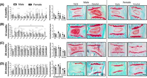
A longitudinal study was conducted to characterize age-, anatomical-, and sex-specific differences in IVD degeneration in two commonly used strains of mice, C57BL/6 and CD-1. Histopathological evaluation of the cervical, thoracic, lumbar, and caudal regions of mice at 6, 12, 20, and 24 months of age was conducted by two blinded observers at each IVD for the nucleus pulposus (NP), annulus fibrosus (AF), and the NP/AF boundary compartments, enabling analysis of scores by tissue compartment, summed scores for each IVD, or averaged scores for each anatomical region.

Results

C57BL/6 mice displayed mild IVD degeneration until 24 months of age; at this point, the lumbar spine demonstrated the most degeneration compared to other regions. Degeneration was detected earlier in the CD-1 mice (20 months of age) in both the thoracic and lumbar spine. In CD-1 mice, moderate to severe degeneration was noted in the cervical spine at all time points assessed. In both strains, age-associated IVD degeneration in the thoracic and lumbar spine was associated with increased histopathological scores in all IVD compartments. In both strains, minimal degeneration was detected in caudal IVDs out to 24 months of age. Both C57BL/6 and CD-1 mice displayed sex-specific differences in the presentation and progression of age-associated IVD degeneration.

Conclusions

These results showed that the progression and severity of age-associated degeneration in mouse models is associated with marked differences based on anatomical region, sex, and strain. This information provides a fundamental baseline characterization for users of mouse models to enable effective and appropriate experimental design, interpretation, and comparison between studies.
Abstract Image

查看原文
分享 分享
微信好友 朋友圈 QQ好友 复制链接
本刊更多论文
对 CD-1 和 C57BL/6 小鼠年龄相关性椎间盘退化的组织病理学比较分析:解剖学和性别差异
椎间盘(IVD)退变是背部疼痛和残疾的主要原因。IVD变性的原因是多因素的,没有疾病修饰治疗。常用小鼠模型研究IVD变性;然而,解剖位置、应变和性别对年龄相关退变进展的影响尚不清楚。一项纵向研究对C57BL/6和CD - 1两种常用小鼠品系的IVD变性进行了年龄、解剖和性别特异性差异的表征。在6、12、20和24月龄时,由两名盲法观察者对小鼠的颈椎、胸椎、腰椎和尾侧区域进行组织病理学评估,分别在每个IVD处对髓核(NP)、纤维环(AF)和NP/AF边界区室进行评估,从而可以按组织区室、每个IVD的总得分或每个解剖区域的平均得分进行分析。C57BL/6小鼠在24月龄前表现为轻度IVD变性;此时,与其他部位相比,腰椎表现出最严重的退变。在CD‐1小鼠(20个月大)中,在胸椎和腰椎的退变更早被检测到。在CD‐1小鼠中,在评估的所有时间点都注意到颈椎中度至重度退变。在这两个品系中,年龄相关的胸椎和腰椎IVD退变与所有IVD区室的组织病理学评分增加相关。在这两个菌株中,在24个月大的尾侧IVDs中检测到最小的变性。C57BL/6和CD - 1小鼠在年龄相关IVD变性的表现和进展方面均表现出性别特异性差异。这些结果表明,小鼠模型中年龄相关变性的进展和严重程度与基于解剖区域、性别和品系的显著差异相关。这些信息为小鼠模型的使用者提供了一个基本的基线特征,以实现有效和适当的实验设计、解释和研究之间的比较。
本文章由计算机程序翻译,如有差异,请以英文原文为准。
求助全文
约1分钟内获得全文 去求助
来源期刊
JOR Spine
JOR Spine ORTHOPEDICS-
CiteScore
6.40
自引率
18.90%
发文量
42
审稿时长
10 weeks
期刊最新文献
The effects of extracellular matrix-degrading enzymes polymorphisms on intervertebral disc degeneration Effect of cigarette smoke exposure and cessation on regional diffusion properties in rat intervertebral discs Pharmacokinetics of PP353, a formulation of linezolid for intervertebral disc administration, in patients with chronic low back pain and Modic change Type 1: A first-in-human, Phase 1b, open-label, single-dose study Preclinical development and characterisation of PP353, a formulation of linezolid for intradiscal administration Melatonin attenuates degenerative disc degression by downregulating DLX5 via the TGF/Smad2/3 pathway in nucleus pulposus cells
×
引用
GB/T 7714-2015
复制
MLA
复制
APA
复制
导出至
BibTeX EndNote RefMan NoteFirst NoteExpress
×
×
提示
您的信息不完整,为了账户安全,请先补充。
现在去补充
×
提示
您因"违规操作"
具体请查看互助需知
我知道了
×
提示
现在去查看 取消
×
提示
确定
0
微信
客服QQ
Book学术公众号 扫码关注我们
反馈
×
意见反馈
请填写您的意见或建议
请填写您的手机或邮箱
已复制链接
已复制链接
快去分享给好友吧!
我知道了
×
扫码分享
扫码分享
Book学术官方微信
Book学术文献互助
Book学术文献互助群
群 号:481959085
Book学术
文献互助 智能选刊 最新文献 互助须知 联系我们:info@booksci.cn
Book学术提供免费学术资源搜索服务,方便国内外学者检索中英文文献。致力于提供最便捷和优质的服务体验。
Copyright © 2023 Book学术 All rights reserved.
ghs 京公网安备 11010802042870号 京ICP备2023020795号-1