Enrica Bassi, Anna Russo, Eugenio Oliboni, Federico Zamboni, Cecilia De Santis, Giancarlo Mansueto, Stefania Montemezzi, Giovanni Foti
{"title":"The role of an artificial intelligence software in clinical senology: a mammography multi-reader study","authors":"Enrica Bassi, Anna Russo, Eugenio Oliboni, Federico Zamboni, Cecilia De Santis, Giancarlo Mansueto, Stefania Montemezzi, Giovanni Foti","doi":"10.1007/s11547-023-01751-1","DOIUrl":null,"url":null,"abstract":"<h3 data-test=\"abstract-sub-heading\">Purpose</h3><p>To evaluate the diagnostic role of a dedicated AI software in detecting anomalous breast findings on mammography and tomosynthesis images in the clinical setting, stand-alone and as aid of four readers.</p><h3 data-test=\"abstract-sub-heading\">Methods</h3><p>A total of 210 patients with complete clinical and radiologic records were retrospectively analyzed. Pathology was used as the reference standard for patients undergoing surgery or biopsy, and a 1-year follow-up was used to confirm no change in the remaining patients.</p><p>The image evaluation was performed by four readers with different levels of experience (a junior and three senior breast radiologists) using a 5-point Likert scale moving from 1 (definitively no cancer) to 5 (definitively cancer).</p><p>The positivity of mammograms was assessed on the presence of any breast lesion (masses, architectural distortions, asymmetries, calcifications), including malignant and benign ones. A multi-reader multi-case analysis was performed. A <i>p</i> value < 0.05 was considered statistically significant.</p><h3 data-test=\"abstract-sub-heading\">Results</h3><p>The stand-alone AI system achieved an accuracy of 71% (69% sensitivity and 73% specificity), which is overall lower than the value achieved by readers without AI. However, with the aid of AI, a significant increase of accuracy (<i>p</i> value = 0.004) and specificity (<i>p</i> value = 0.04) was achieved for the less experienced radiologist and a senior one.</p><h3 data-test=\"abstract-sub-heading\">Conclusion</h3><p>The use of AI software as a second reader for breast lesions assessment could play a crucial role in the clinical setting, by increasing sensitivity and specificity, especially for less experienced radiologists.</p>","PeriodicalId":501689,"journal":{"name":"La radiologia medica","volume":"84 1","pages":""},"PeriodicalIF":0.0000,"publicationDate":"2023-12-11","publicationTypes":"Journal Article","fieldsOfStudy":null,"isOpenAccess":false,"openAccessPdf":"","citationCount":"0","resultStr":null,"platform":"Semanticscholar","paperid":null,"PeriodicalName":"La radiologia medica","FirstCategoryId":"1085","ListUrlMain":"https://doi.org/10.1007/s11547-023-01751-1","RegionNum":0,"RegionCategory":null,"ArticlePicture":[],"TitleCN":null,"AbstractTextCN":null,"PMCID":null,"EPubDate":"","PubModel":"","JCR":"","JCRName":"","Score":null,"Total":0}
引用次数: 0
Abstract
Purpose
To evaluate the diagnostic role of a dedicated AI software in detecting anomalous breast findings on mammography and tomosynthesis images in the clinical setting, stand-alone and as aid of four readers.
Methods
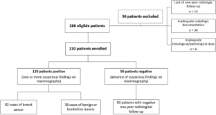
A total of 210 patients with complete clinical and radiologic records were retrospectively analyzed. Pathology was used as the reference standard for patients undergoing surgery or biopsy, and a 1-year follow-up was used to confirm no change in the remaining patients.
The image evaluation was performed by four readers with different levels of experience (a junior and three senior breast radiologists) using a 5-point Likert scale moving from 1 (definitively no cancer) to 5 (definitively cancer).
The positivity of mammograms was assessed on the presence of any breast lesion (masses, architectural distortions, asymmetries, calcifications), including malignant and benign ones. A multi-reader multi-case analysis was performed. A p value < 0.05 was considered statistically significant.
Results
The stand-alone AI system achieved an accuracy of 71% (69% sensitivity and 73% specificity), which is overall lower than the value achieved by readers without AI. However, with the aid of AI, a significant increase of accuracy (p value = 0.004) and specificity (p value = 0.04) was achieved for the less experienced radiologist and a senior one.
Conclusion
The use of AI software as a second reader for breast lesions assessment could play a crucial role in the clinical setting, by increasing sensitivity and specificity, especially for less experienced radiologists.


 分享
分享

 求助内容:
求助内容: 应助结果提醒方式:
 扫码关注我们
