{"title":"Epichordal vertebral column formation in Xenopus laevis","authors":"Yu Takahashi, Ryota Wakabayashi, Satoshi Kitajima, Hideho Uchiyama","doi":"10.1002/jmor.21664","DOIUrl":null,"url":null,"abstract":"<p>Although <i>Xenopus Laevis</i> is the most widely used model amphibian, skeletal development of its vertebral column has not been well illustrated so far. The mode of vertebral column development in anurans has been classified into two modes: perichordal and epichordal. <i>Xenopus</i> vertebral column formation is believed to follow the epichordal mode, but this aspect has been underemphasized, and illustrative examples are currently unavailable to the scientific community. This study documents the entire process of vertebral column formation in <i>X. laevis</i>, from the initial neural arch formation to the completion of metamorphosis. These images reveal that the neural arch arises from the dorsal lamina and lateral pedicle primordia, with no strict adherence to an anteroposterior sequence. Unlike other species, <i>Xenopus</i> centrum primordia exclusively form at the expanded ventral margins of neural arches, rather than from the cartilaginous layer surrounding the notochord. These paired centrum primordia then fuse at the ventral midline, dorsal to the notochord, and subsequently the notochord degenerates. This mode of centrum formation differs from the traditional epichordal mode, indicating that <i>Xenopus</i> might have lost the ability to form a cartilaginous layer around the notochord. Instead, the neural arch's ventral margin appears to have evolved to incorporate centrum precursor cells at its base, thereby forming a centrum-like structure compensating for the absence of a true centrum. It is widely accepted that postsacral vertebrae lack centra, only possessing neural arches, and eventually fuse with the hypochord to form the urostyle. However, we have shown that the paired ventral ends of the postsacral vertebrae also fuse at the midline to form a centrum-like structure. This process might extend to the trunk region during centrum formation. In addition to these findings, we offer evolutionary insights into the reasons why <i>Xenopus</i> retains centrum primordia at the base of neural arches.</p>","PeriodicalId":16528,"journal":{"name":"Journal of Morphology","volume":"285 2","pages":""},"PeriodicalIF":1.4000,"publicationDate":"2023-12-18","publicationTypes":"Journal Article","fieldsOfStudy":null,"isOpenAccess":false,"openAccessPdf":"","citationCount":"0","resultStr":null,"platform":"Semanticscholar","paperid":null,"PeriodicalName":"Journal of Morphology","FirstCategoryId":"3","ListUrlMain":"https://onlinelibrary.wiley.com/doi/10.1002/jmor.21664","RegionNum":4,"RegionCategory":"医学","ArticlePicture":[],"TitleCN":null,"AbstractTextCN":null,"PMCID":null,"EPubDate":"","PubModel":"","JCR":"Q2","JCRName":"ANATOMY & MORPHOLOGY","Score":null,"Total":0}
引用次数: 0
Abstract
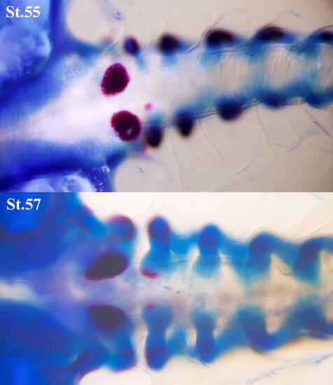
Although Xenopus Laevis is the most widely used model amphibian, skeletal development of its vertebral column has not been well illustrated so far. The mode of vertebral column development in anurans has been classified into two modes: perichordal and epichordal. Xenopus vertebral column formation is believed to follow the epichordal mode, but this aspect has been underemphasized, and illustrative examples are currently unavailable to the scientific community. This study documents the entire process of vertebral column formation in X. laevis, from the initial neural arch formation to the completion of metamorphosis. These images reveal that the neural arch arises from the dorsal lamina and lateral pedicle primordia, with no strict adherence to an anteroposterior sequence. Unlike other species, Xenopus centrum primordia exclusively form at the expanded ventral margins of neural arches, rather than from the cartilaginous layer surrounding the notochord. These paired centrum primordia then fuse at the ventral midline, dorsal to the notochord, and subsequently the notochord degenerates. This mode of centrum formation differs from the traditional epichordal mode, indicating that Xenopus might have lost the ability to form a cartilaginous layer around the notochord. Instead, the neural arch's ventral margin appears to have evolved to incorporate centrum precursor cells at its base, thereby forming a centrum-like structure compensating for the absence of a true centrum. It is widely accepted that postsacral vertebrae lack centra, only possessing neural arches, and eventually fuse with the hypochord to form the urostyle. However, we have shown that the paired ventral ends of the postsacral vertebrae also fuse at the midline to form a centrum-like structure. This process might extend to the trunk region during centrum formation. In addition to these findings, we offer evolutionary insights into the reasons why Xenopus retains centrum primordia at the base of neural arches.
期刊介绍:
The Journal of Morphology welcomes articles of original research in cytology, protozoology, embryology, and general morphology. Articles generally should not exceed 35 printed pages. Preliminary notices or articles of a purely descriptive morphological or taxonomic nature are not included. No paper which has already been published will be accepted, nor will simultaneous publications elsewhere be allowed.
The Journal of Morphology publishes research in functional, comparative, evolutionary and developmental morphology from vertebrates and invertebrates. Human and veterinary anatomy or paleontology are considered when an explicit connection to neontological animal morphology is presented, and the paper contains relevant information for the community of animal morphologists. Based on our long tradition, we continue to seek publishing the best papers in animal morphology.


 分享
分享
 求助内容:
求助内容: 应助结果提醒方式:
 扫码关注我们
