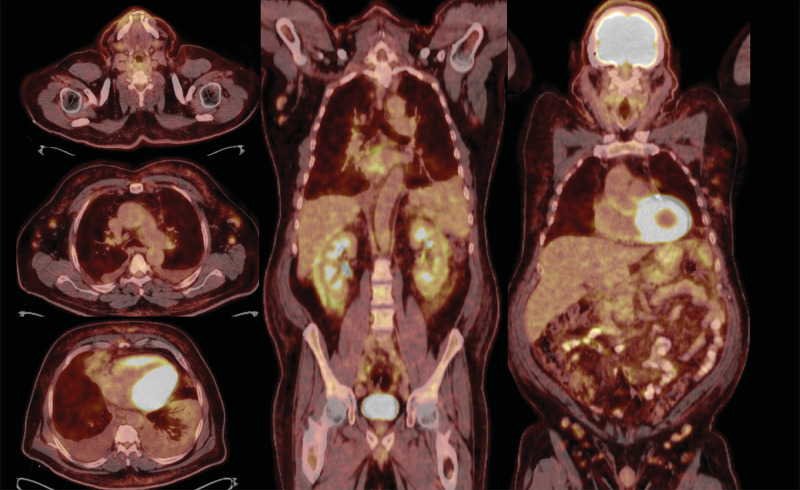
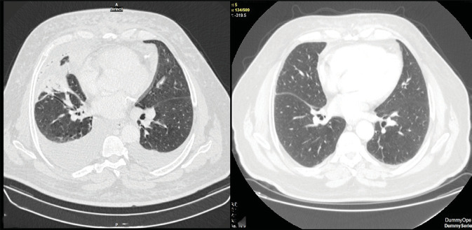
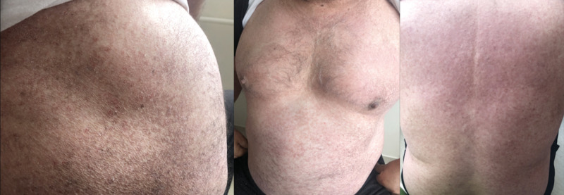
{"title":"A patient with widespread skin lesions presenting with massive pleural effusion.","authors":"Aslıhan Gürün Kaya, Pervin Topçuoğlu, Ayten Kayı Cangır, Nihal Kundakçı, Işınsu Kuzu, Aylin Okçu Heper, Demet Karnak","doi":"10.5578/tt.20239610","DOIUrl":null,"url":null,"abstract":"<p><p>Mycosis fungoides is the most commonly seen type of cutaneous T-cell lymphoproliferative disease. While mycosis fungoides is linked to an increased risk of developing secondary malignancies, the occurrence of B-cell-originated disease in association with it is exceedingly rare. A 66-year-old male with persistent papillomatous skin eruption was admitted due to dyspnea. Chest X-ray, positron emission tomography, and chest computed tomography revealed axillary and mediastinal lymph node enlargement and right lower pulmonary lobe infiltration along with right-sided massive pleural effusion. Histological and immunohistochemical findings of pleural biopsy and axillary lymph nodes suggested a diagnosis of pulmonary extranodal marginal zone lymphoma. Skin biopsies from the abdomen, chest, and legs revealed CD4/CD8 double-positive patch stage of mycosis fungoides. After completing six cycles of chemotherapy, complete remission of lymphoma was achieved, with the skin eruptions remaining unchanged. Herein, the authors present a unique case of concomitant diagnoses of mycosis fungoides and marginal zone B-cell lymphoma of the respiratory system to emphasize the importance of careful evaluation of each finding.</p>","PeriodicalId":45521,"journal":{"name":"Tuberkuloz ve Toraks-Tuberculosis and Thorax","volume":"71 4","pages":"420-427"},"PeriodicalIF":0.7000,"publicationDate":"2023-12-01","publicationTypes":"Journal Article","fieldsOfStudy":null,"isOpenAccess":false,"openAccessPdf":"https://www.ncbi.nlm.nih.gov/pmc/articles/PMC11390065/pdf/","citationCount":"0","resultStr":null,"platform":"Semanticscholar","paperid":null,"PeriodicalName":"Tuberkuloz ve Toraks-Tuberculosis and Thorax","FirstCategoryId":"1085","ListUrlMain":"https://doi.org/10.5578/tt.20239610","RegionNum":0,"RegionCategory":null,"ArticlePicture":[],"TitleCN":null,"AbstractTextCN":null,"PMCID":null,"EPubDate":"","PubModel":"","JCR":"Q4","JCRName":"RESPIRATORY SYSTEM","Score":null,"Total":0}
引用次数: 0
Abstract
Mycosis fungoides is the most commonly seen type of cutaneous T-cell lymphoproliferative disease. While mycosis fungoides is linked to an increased risk of developing secondary malignancies, the occurrence of B-cell-originated disease in association with it is exceedingly rare. A 66-year-old male with persistent papillomatous skin eruption was admitted due to dyspnea. Chest X-ray, positron emission tomography, and chest computed tomography revealed axillary and mediastinal lymph node enlargement and right lower pulmonary lobe infiltration along with right-sided massive pleural effusion. Histological and immunohistochemical findings of pleural biopsy and axillary lymph nodes suggested a diagnosis of pulmonary extranodal marginal zone lymphoma. Skin biopsies from the abdomen, chest, and legs revealed CD4/CD8 double-positive patch stage of mycosis fungoides. After completing six cycles of chemotherapy, complete remission of lymphoma was achieved, with the skin eruptions remaining unchanged. Herein, the authors present a unique case of concomitant diagnoses of mycosis fungoides and marginal zone B-cell lymphoma of the respiratory system to emphasize the importance of careful evaluation of each finding.
真菌病是最常见的皮肤 T 细胞淋巴组织增生性疾病。虽然放线菌病与继发性恶性肿瘤的发病风险增加有关,但与之相关的B细胞源性疾病却极为罕见。一名66岁的男性因呼吸困难入院,他患有持续性乳头状皮肤糜烂。胸部 X 光、正电子发射断层扫描和胸部计算机断层扫描显示腋窝和纵隔淋巴结肿大、右下肺叶浸润以及右侧大量胸腔积液。胸膜活检和腋窝淋巴结的组织学和免疫组化结果表明,诊断为肺外缘区淋巴瘤。腹部、胸部和腿部的皮肤活检显示,CD4/CD8双阳性斑块处于真菌病阶段。在完成六个周期的化疗后,淋巴瘤完全缓解,皮肤糜烂的情况没有改变。在此,作者介绍了一例同时诊断为真菌病和呼吸系统边缘区B细胞淋巴瘤的独特病例,以强调对每项发现进行仔细评估的重要性。


 分享
分享
 求助内容:
求助内容: 应助结果提醒方式:
 扫码关注我们
