Madan M Maddali, Robert H Anderson, Salim N Al Maskari, Faiza Al Kindi, Hamood N Al Kindi
{"title":"The Sinus Venosus Veno-Venous Bridge: Not a septal defect.","authors":"Madan M Maddali, Robert H Anderson, Salim N Al Maskari, Faiza Al Kindi, Hamood N Al Kindi","doi":"10.18295/squmj.12.2023.075","DOIUrl":null,"url":null,"abstract":"<p><p>This review provides an update on the morphology of the sinus venosus defect. It was earlier believed that a 'common wall' separated the right pulmonary veins from the superior caval vein. In the sinus venosus defects, this wall was absent. Current evidence shows that the superior rim of the oval fossa, rather than forming a second septum or representing a common wall, is an infolding between the walls of the caval veins and the right pulmonary veins. The sinus venosus defect is caused by the anomalous connection of one or more pulmonary veins to a systemic vein. However, the pulmonary vein(s) retain their left atrial connections, leading to a veno-venous bridge that allows interatrial shunting outside the oval fossa. True atrial septal defects are located within the oval fossa or in the anteo-inferior buttress, while sinus venosus defects, ostium defects and coronary sinus defects are morphologically distinct from them.</p>","PeriodicalId":22083,"journal":{"name":"Sultan Qaboos University Medical Journal","volume":"23 Spec Iss","pages":"5-9"},"PeriodicalIF":0.0000,"publicationDate":"2023-12-01","publicationTypes":"Journal Article","fieldsOfStudy":null,"isOpenAccess":false,"openAccessPdf":"https://www.ncbi.nlm.nih.gov/pmc/articles/PMC10754305/pdf/","citationCount":"0","resultStr":null,"platform":"Semanticscholar","paperid":null,"PeriodicalName":"Sultan Qaboos University Medical Journal","FirstCategoryId":"1085","ListUrlMain":"https://doi.org/10.18295/squmj.12.2023.075","RegionNum":0,"RegionCategory":null,"ArticlePicture":[],"TitleCN":null,"AbstractTextCN":null,"PMCID":null,"EPubDate":"2023/11/30 0:00:00","PubModel":"Epub","JCR":"Q3","JCRName":"Medicine","Score":null,"Total":0}
引用次数: 0
Abstract
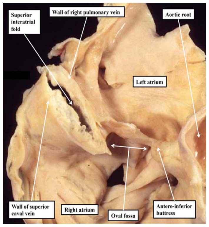
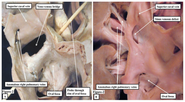
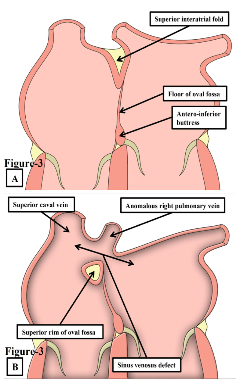
This review provides an update on the morphology of the sinus venosus defect. It was earlier believed that a 'common wall' separated the right pulmonary veins from the superior caval vein. In the sinus venosus defects, this wall was absent. Current evidence shows that the superior rim of the oval fossa, rather than forming a second septum or representing a common wall, is an infolding between the walls of the caval veins and the right pulmonary veins. The sinus venosus defect is caused by the anomalous connection of one or more pulmonary veins to a systemic vein. However, the pulmonary vein(s) retain their left atrial connections, leading to a veno-venous bridge that allows interatrial shunting outside the oval fossa. True atrial septal defects are located within the oval fossa or in the anteo-inferior buttress, while sinus venosus defects, ostium defects and coronary sinus defects are morphologically distinct from them.


 分享
分享
 求助内容:
求助内容: 应助结果提醒方式:
 扫码关注我们
