Yameng Zhang, Xinping Qi, Weitao Li, Min Wan, Xue Ning, Jin Hu
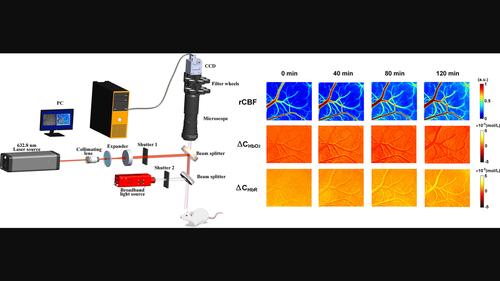
{"title":"Research on the classification of early-stage brain edema by combining intrinsic optical signal imaging and laser speckle contrast imaging","authors":"Yameng Zhang, Xinping Qi, Weitao Li, Min Wan, Xue Ning, Jin Hu","doi":"10.1002/jbio.202300394","DOIUrl":null,"url":null,"abstract":"<p>The early detection and pathological classification of brain edema are very important for symptomatic treatment. The dual-optical imaging system (DOIS) consists of intrinsic optical signal imaging (IOSI) and laser speckle contrast imaging (LSCI), which can acquire cerebral hemodynamic parameters of mice in real-time, including changes of oxygenated hemoglobin concentration (<math>\n <mrow>\n <mi>Δ</mi>\n <msub>\n <mi>C</mi>\n <msub>\n <mi>HbO</mi>\n <mn>2</mn>\n </msub>\n </msub>\n </mrow></math>), deoxyhemoglobin concentration (Δ<i>C</i><sub>HbR</sub>) and relative cerebral blood flow (rCBF) within the field of view. The slope sum of <math>\n <mrow>\n <mi>Δ</mi>\n <msub>\n <mi>C</mi>\n <msub>\n <mi>HbO</mi>\n <mn>2</mn>\n </msub>\n </msub>\n </mrow></math>, Δ<i>C</i><sub>HbR</sub> and rCBF was proposed to classify vasogenic edema (VE) and cytotoxic edema (CE). The slope sum values in the VE and CE group remain statistically different and the classification results provide higher accuracy of more than 93% for early brain edema detection. In conclusion, the differences of hemodynamic parameters between VE and CE in the early stage were revealed and the method helps in the classification of early brain edema.</p>","PeriodicalId":184,"journal":{"name":"Journal of Biophotonics","volume":"17 3","pages":""},"PeriodicalIF":2.0000,"publicationDate":"2024-01-02","publicationTypes":"Journal Article","fieldsOfStudy":null,"isOpenAccess":false,"openAccessPdf":"","citationCount":"0","resultStr":null,"platform":"Semanticscholar","paperid":null,"PeriodicalName":"Journal of Biophotonics","FirstCategoryId":"101","ListUrlMain":"https://onlinelibrary.wiley.com/doi/10.1002/jbio.202300394","RegionNum":3,"RegionCategory":"物理与天体物理","ArticlePicture":[],"TitleCN":null,"AbstractTextCN":null,"PMCID":null,"EPubDate":"","PubModel":"","JCR":"Q3","JCRName":"BIOCHEMICAL RESEARCH METHODS","Score":null,"Total":0}
引用次数: 0
Abstract
The early detection and pathological classification of brain edema are very important for symptomatic treatment. The dual-optical imaging system (DOIS) consists of intrinsic optical signal imaging (IOSI) and laser speckle contrast imaging (LSCI), which can acquire cerebral hemodynamic parameters of mice in real-time, including changes of oxygenated hemoglobin concentration (), deoxyhemoglobin concentration (ΔCHbR) and relative cerebral blood flow (rCBF) within the field of view. The slope sum of , ΔCHbR and rCBF was proposed to classify vasogenic edema (VE) and cytotoxic edema (CE). The slope sum values in the VE and CE group remain statistically different and the classification results provide higher accuracy of more than 93% for early brain edema detection. In conclusion, the differences of hemodynamic parameters between VE and CE in the early stage were revealed and the method helps in the classification of early brain edema.
期刊介绍:
The first international journal dedicated to publishing reviews and original articles from this exciting field, the Journal of Biophotonics covers the broad range of research on interactions between light and biological material. The journal offers a platform where the physicist communicates with the biologist and where the clinical practitioner learns about the latest tools for the diagnosis of diseases. As such, the journal is highly interdisciplinary, publishing cutting edge research in the fields of life sciences, medicine, physics, chemistry, and engineering. The coverage extends from fundamental research to specific developments, while also including the latest applications.


 分享
分享
 求助内容:
求助内容: 应助结果提醒方式:
 扫码关注我们
