Janavi Subramani, Niharika Patlolla, Rajani Battu, Taslimarif Saiyed, Rajarshi Pal
{"title":"Generation and characterization of retinal pigment epithelium from patient iPSC line to model oculocutaneous albinism (OCA)1A disease","authors":"Janavi Subramani, Niharika Patlolla, Rajani Battu, Taslimarif Saiyed, Rajarshi Pal","doi":"10.1007/s12038-023-00406-7","DOIUrl":null,"url":null,"abstract":"<p>Oculocutaneous albinism (OCA) is characterized by reduced melanin biosynthesis affecting the retina, thus impairing visual function. The disease pathology of OCA is poorly understood at the cellular level due to unavailability of suitable biological model systems. This study aimed to develop a disease-specific <i>in vitro</i> model for OCA type 1A, the most severe form caused by <i>TYR</i> (tyrosinase) gene mutations, using retinal pigment epithelium (RPE) differentiated from patient-derived human-induced pluripotent stem cells (hiPSCs). A comparative study between healthy and OCA1A RPE cells revealed that while healthy RPE cells exhibited timely onest of pigmentation during differentiation, OCA1A RPE cells failed to pigment even after an extended culture period. This observation was validated by ultrastructural studies using electron microscopy, hinting at melanosome-specific defects. Immunocytochemistry demonstrated abnormal expression patterns of melanogenesis-specific protein markers in OCA1A RPE cells, indicating reduced or absence of melanin synthesis. Next, a quantitative assay was performed to confirm the absence of melanin production in OCA1A RPE cells. Tyrosinase assay showed no activity in OCA1A compared with healthy RPE, suggesting non-functionality of <i>TYR</i>, further corroborated by western blot analysis showing complete absence of the protein. Gene expression by RNA sequencing of healthy and OCA1A RPE cells uncovered differential gene expression associated with lens development, visual perception, transmembrane transporter activity, and key signaling pathways. This disease-in-a-dish model of OCA1A provides an excellent platform to understand disease mechanism, identify potential therapeutic targets, and facilitate gene therapy or gene correction.</p>","PeriodicalId":15171,"journal":{"name":"Journal of Biosciences","volume":"12 1","pages":""},"PeriodicalIF":1.9000,"publicationDate":"2024-01-20","publicationTypes":"Journal Article","fieldsOfStudy":null,"isOpenAccess":false,"openAccessPdf":"","citationCount":"0","resultStr":null,"platform":"Semanticscholar","paperid":null,"PeriodicalName":"Journal of Biosciences","FirstCategoryId":"99","ListUrlMain":"https://doi.org/10.1007/s12038-023-00406-7","RegionNum":4,"RegionCategory":"生物学","ArticlePicture":[],"TitleCN":null,"AbstractTextCN":null,"PMCID":null,"EPubDate":"","PubModel":"","JCR":"Q2","JCRName":"BIOLOGY","Score":null,"Total":0}
引用次数: 0
Abstract
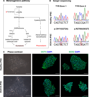
Oculocutaneous albinism (OCA) is characterized by reduced melanin biosynthesis affecting the retina, thus impairing visual function. The disease pathology of OCA is poorly understood at the cellular level due to unavailability of suitable biological model systems. This study aimed to develop a disease-specific in vitro model for OCA type 1A, the most severe form caused by TYR (tyrosinase) gene mutations, using retinal pigment epithelium (RPE) differentiated from patient-derived human-induced pluripotent stem cells (hiPSCs). A comparative study between healthy and OCA1A RPE cells revealed that while healthy RPE cells exhibited timely onest of pigmentation during differentiation, OCA1A RPE cells failed to pigment even after an extended culture period. This observation was validated by ultrastructural studies using electron microscopy, hinting at melanosome-specific defects. Immunocytochemistry demonstrated abnormal expression patterns of melanogenesis-specific protein markers in OCA1A RPE cells, indicating reduced or absence of melanin synthesis. Next, a quantitative assay was performed to confirm the absence of melanin production in OCA1A RPE cells. Tyrosinase assay showed no activity in OCA1A compared with healthy RPE, suggesting non-functionality of TYR, further corroborated by western blot analysis showing complete absence of the protein. Gene expression by RNA sequencing of healthy and OCA1A RPE cells uncovered differential gene expression associated with lens development, visual perception, transmembrane transporter activity, and key signaling pathways. This disease-in-a-dish model of OCA1A provides an excellent platform to understand disease mechanism, identify potential therapeutic targets, and facilitate gene therapy or gene correction.
期刊介绍:
The Journal of Biosciences is a quarterly journal published by the Indian Academy of Sciences, Bangalore. It covers all areas of Biology and is the premier journal in the country within its scope. It is indexed in Current Contents and other standard Biological and Medical databases. The Journal of Biosciences began in 1934 as the Proceedings of the Indian Academy of Sciences (Section B). This continued until 1978 when it was split into three parts : Proceedings-Animal Sciences, Proceedings-Plant Sciences and Proceedings-Experimental Biology. Proceedings-Experimental Biology was renamed Journal of Biosciences in 1979; and in 1991, Proceedings-Animal Sciences and Proceedings-Plant Sciences merged with it.


 分享
分享
 求助内容:
求助内容: 应助结果提醒方式:
 扫码关注我们
