Aline Knab, Ayad G. Anwer, Bernadette Pedersen, Shannon Handley, Abhilash Goud Marupally, Abbas Habibalahi, Ewa M. Goldys
{"title":"Towards label-free non-invasive autofluorescence multispectral imaging for melanoma diagnosis","authors":"Aline Knab, Ayad G. Anwer, Bernadette Pedersen, Shannon Handley, Abhilash Goud Marupally, Abbas Habibalahi, Ewa M. Goldys","doi":"10.1002/jbio.202300402","DOIUrl":null,"url":null,"abstract":"<p>This study focuses on the use of cellular autofluorescence which visualizes the cell metabolism by monitoring endogenous fluorophores including NAD(P)H and flavins. It explores the potential of multispectral imaging of native fluorophores in melanoma diagnostics using excitation wavelengths ranging from 340 nm to 510 nm and emission wavelengths above 391 nm. Cultured immortalized cells are utilized to compare the autofluorescent signatures of two melanoma cell lines to one fibroblast cell line. Feature analysis identifies the most significant and least correlated features for differentiating the cells. The investigation successfully applies this analysis to pre-processed, noise-removed images and original background-corrupted data. Furthermore, the applicability of distinguishing melanomas and healthy fibroblasts based on their autofluorescent characteristics is validated using the same evaluation technique on patient cells. Additionally, the study tentatively maps the detected features to underlying biological processes. This research demonstrates the potential of cellular autofluorescence as a promising tool for melanoma diagnostics.</p>","PeriodicalId":184,"journal":{"name":"Journal of Biophotonics","volume":"17 4","pages":""},"PeriodicalIF":2.0000,"publicationDate":"2024-01-21","publicationTypes":"Journal Article","fieldsOfStudy":null,"isOpenAccess":false,"openAccessPdf":"https://onlinelibrary.wiley.com/doi/epdf/10.1002/jbio.202300402","citationCount":"0","resultStr":null,"platform":"Semanticscholar","paperid":null,"PeriodicalName":"Journal of Biophotonics","FirstCategoryId":"101","ListUrlMain":"https://onlinelibrary.wiley.com/doi/10.1002/jbio.202300402","RegionNum":3,"RegionCategory":"物理与天体物理","ArticlePicture":[],"TitleCN":null,"AbstractTextCN":null,"PMCID":null,"EPubDate":"","PubModel":"","JCR":"Q3","JCRName":"BIOCHEMICAL RESEARCH METHODS","Score":null,"Total":0}
引用次数: 0
Abstract
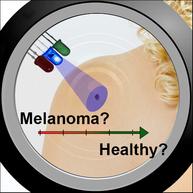
This study focuses on the use of cellular autofluorescence which visualizes the cell metabolism by monitoring endogenous fluorophores including NAD(P)H and flavins. It explores the potential of multispectral imaging of native fluorophores in melanoma diagnostics using excitation wavelengths ranging from 340 nm to 510 nm and emission wavelengths above 391 nm. Cultured immortalized cells are utilized to compare the autofluorescent signatures of two melanoma cell lines to one fibroblast cell line. Feature analysis identifies the most significant and least correlated features for differentiating the cells. The investigation successfully applies this analysis to pre-processed, noise-removed images and original background-corrupted data. Furthermore, the applicability of distinguishing melanomas and healthy fibroblasts based on their autofluorescent characteristics is validated using the same evaluation technique on patient cells. Additionally, the study tentatively maps the detected features to underlying biological processes. This research demonstrates the potential of cellular autofluorescence as a promising tool for melanoma diagnostics.
期刊介绍:
The first international journal dedicated to publishing reviews and original articles from this exciting field, the Journal of Biophotonics covers the broad range of research on interactions between light and biological material. The journal offers a platform where the physicist communicates with the biologist and where the clinical practitioner learns about the latest tools for the diagnosis of diseases. As such, the journal is highly interdisciplinary, publishing cutting edge research in the fields of life sciences, medicine, physics, chemistry, and engineering. The coverage extends from fundamental research to specific developments, while also including the latest applications.


 分享
分享
 求助内容:
求助内容: 应助结果提醒方式:
 扫码关注我们
