Detection of acute and early-delayed radiation-induced changes in the white matter of the rat brain based on numerical processing of optical coherence tomography data
Ksenia Achkasova, Liudmila Kukhnina, Alexander Moiseev, Elena Kiseleva, Alexandra Bogomolova, Maria Loginova, Natalia Gladkova
{"title":"Detection of acute and early-delayed radiation-induced changes in the white matter of the rat brain based on numerical processing of optical coherence tomography data","authors":"Ksenia Achkasova, Liudmila Kukhnina, Alexander Moiseev, Elena Kiseleva, Alexandra Bogomolova, Maria Loginova, Natalia Gladkova","doi":"10.1002/jbio.202300458","DOIUrl":null,"url":null,"abstract":"<p>Detection of radiation-induced changes of the brain white matter is important for brain neoplasms repeated surgery. We investigated the influence of irradiation on the scattering properties of the white matter using optical coherence tomography (OCT). Healthy Wistar rats undergone the irradiation of the brain right hemisphere. At seven time points from the irradiation procedure (2–14 weeks), an ex vivo OCT study was performed with subsequent calculation of attenuation coefficient values in the corpus callosum followed by immunohistochemical analysis. As a result, we discovered acute and early-delayed changes characterized by the edema of different severity, accompanied by a statistically significant decrease in attenuation coefficient values. In particular, these changes were found at 2 weeks after irradiation in the irradiated hemisphere, while at 6- and 12-week time points they affected both irradiated and contralateral hemisphere. Thus, radiation-induced changes occurring in white matter during the first 3 months after irradiation can be detected by OCT.</p>","PeriodicalId":184,"journal":{"name":"Journal of Biophotonics","volume":null,"pages":null},"PeriodicalIF":2.0000,"publicationDate":"2024-01-22","publicationTypes":"Journal Article","fieldsOfStudy":null,"isOpenAccess":false,"openAccessPdf":"","citationCount":"0","resultStr":null,"platform":"Semanticscholar","paperid":null,"PeriodicalName":"Journal of Biophotonics","FirstCategoryId":"101","ListUrlMain":"https://onlinelibrary.wiley.com/doi/10.1002/jbio.202300458","RegionNum":3,"RegionCategory":"物理与天体物理","ArticlePicture":[],"TitleCN":null,"AbstractTextCN":null,"PMCID":null,"EPubDate":"","PubModel":"","JCR":"Q3","JCRName":"BIOCHEMICAL RESEARCH METHODS","Score":null,"Total":0}
引用次数: 0
Abstract
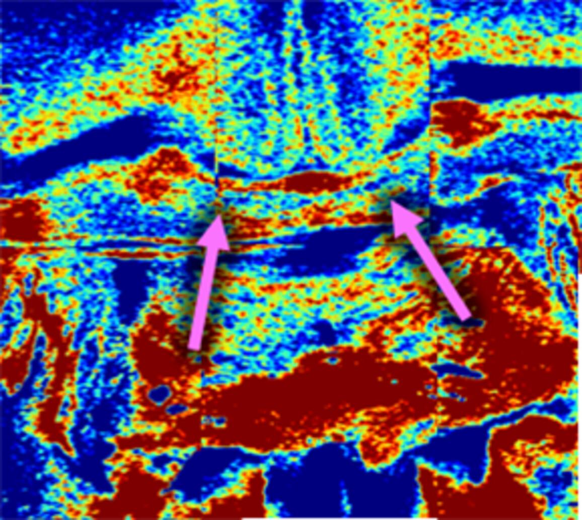
Detection of radiation-induced changes of the brain white matter is important for brain neoplasms repeated surgery. We investigated the influence of irradiation on the scattering properties of the white matter using optical coherence tomography (OCT). Healthy Wistar rats undergone the irradiation of the brain right hemisphere. At seven time points from the irradiation procedure (2–14 weeks), an ex vivo OCT study was performed with subsequent calculation of attenuation coefficient values in the corpus callosum followed by immunohistochemical analysis. As a result, we discovered acute and early-delayed changes characterized by the edema of different severity, accompanied by a statistically significant decrease in attenuation coefficient values. In particular, these changes were found at 2 weeks after irradiation in the irradiated hemisphere, while at 6- and 12-week time points they affected both irradiated and contralateral hemisphere. Thus, radiation-induced changes occurring in white matter during the first 3 months after irradiation can be detected by OCT.
期刊介绍:
The first international journal dedicated to publishing reviews and original articles from this exciting field, the Journal of Biophotonics covers the broad range of research on interactions between light and biological material. The journal offers a platform where the physicist communicates with the biologist and where the clinical practitioner learns about the latest tools for the diagnosis of diseases. As such, the journal is highly interdisciplinary, publishing cutting edge research in the fields of life sciences, medicine, physics, chemistry, and engineering. The coverage extends from fundamental research to specific developments, while also including the latest applications.


 分享
分享
 求助内容:
求助内容: 应助结果提醒方式:
 扫码关注我们
