Sönke Peters, Fernando Bueno Neves, Monika Huhndorf, Friederike Gärtner, Klarissa Stürner, Olav Jansen, Mona Salehi Ravesh
{"title":"Detection of Spinal Cord Multiple Sclerosis Lesions Using a 3D-PSIR Sequence at 1.5 T.","authors":"Sönke Peters, Fernando Bueno Neves, Monika Huhndorf, Friederike Gärtner, Klarissa Stürner, Olav Jansen, Mona Salehi Ravesh","doi":"10.1007/s00062-023-01376-x","DOIUrl":null,"url":null,"abstract":"<p><strong>Purpose: </strong>Multiple sclerosis (MS) is a prevalent autoimmune inflammatory disease. Besides cerebral manifestations, an affection of the spinal cord is typical; however, imaging of the spinal cord is difficult due to its anatomy. The aim of this study was to assess the diagnostic value of a 3D PSIR pulse sequencing at a 1.5 T magnetic field strength for both the cervical and thoracic spinal cord.</p><p><strong>Methods: </strong>Phase sensitive inversion recovery (PSIR), short tau inversion recovery (STIR) and T<sub>2</sub>-weighted (T<sub>2</sub>-w) images of the spinal cord of 50 patients were separately evaluated by three radiologists concerning the number and location of MS lesions. Furthermore, lesion to cord contrast ratios were determined for the cervical and thoracic spinal cord.</p><p><strong>Results: </strong>Of the lesions 54.81% were located in the cervical spinal cord, 42.26% in the thoracic spinal cord and 2.93% in the conus medullaris. The PSIR images showed a higher sensitivity for lesion detection in the cervical and thoracic spinal cord (77.10% and 72.61%, respectively) compared to the STIR images (58.63% and 59.10%, respectively) and the T<sub>2</sub>-w images (59.95% and 59.52%, respectively). The average lesion to cord contrast ratio was significantly higher in the PSIR images compared to the STIR images (p < 0.001) and the T<sub>2</sub>-w images (p < 0.001).</p><p><strong>Conclusion: </strong>Evaluation of the spinal cord with a 3D PSIR sequence at a magnetic field strength of 1.5 T is feasible with a high sensitivity for the detection of spinal MS lesions for the cervical as well as the thoracic segments. In combination with other pulse sequences it might become a valuable addition in an advanced imaging protocol.</p>","PeriodicalId":49298,"journal":{"name":"Clinical Neuroradiology","volume":" ","pages":"403-410"},"PeriodicalIF":2.6000,"publicationDate":"2024-06-01","publicationTypes":"Journal Article","fieldsOfStudy":null,"isOpenAccess":false,"openAccessPdf":"https://www.ncbi.nlm.nih.gov/pmc/articles/PMC11130041/pdf/","citationCount":"0","resultStr":null,"platform":"Semanticscholar","paperid":null,"PeriodicalName":"Clinical Neuroradiology","FirstCategoryId":"3","ListUrlMain":"https://doi.org/10.1007/s00062-023-01376-x","RegionNum":3,"RegionCategory":"医学","ArticlePicture":[],"TitleCN":null,"AbstractTextCN":null,"PMCID":null,"EPubDate":"2024/1/30 0:00:00","PubModel":"Epub","JCR":"Q2","JCRName":"CLINICAL NEUROLOGY","Score":null,"Total":0}
引用次数: 0
Abstract
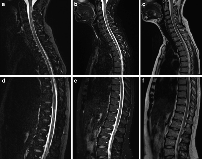
Purpose: Multiple sclerosis (MS) is a prevalent autoimmune inflammatory disease. Besides cerebral manifestations, an affection of the spinal cord is typical; however, imaging of the spinal cord is difficult due to its anatomy. The aim of this study was to assess the diagnostic value of a 3D PSIR pulse sequencing at a 1.5 T magnetic field strength for both the cervical and thoracic spinal cord.
Methods: Phase sensitive inversion recovery (PSIR), short tau inversion recovery (STIR) and T2-weighted (T2-w) images of the spinal cord of 50 patients were separately evaluated by three radiologists concerning the number and location of MS lesions. Furthermore, lesion to cord contrast ratios were determined for the cervical and thoracic spinal cord.
Results: Of the lesions 54.81% were located in the cervical spinal cord, 42.26% in the thoracic spinal cord and 2.93% in the conus medullaris. The PSIR images showed a higher sensitivity for lesion detection in the cervical and thoracic spinal cord (77.10% and 72.61%, respectively) compared to the STIR images (58.63% and 59.10%, respectively) and the T2-w images (59.95% and 59.52%, respectively). The average lesion to cord contrast ratio was significantly higher in the PSIR images compared to the STIR images (p < 0.001) and the T2-w images (p < 0.001).
Conclusion: Evaluation of the spinal cord with a 3D PSIR sequence at a magnetic field strength of 1.5 T is feasible with a high sensitivity for the detection of spinal MS lesions for the cervical as well as the thoracic segments. In combination with other pulse sequences it might become a valuable addition in an advanced imaging protocol.
期刊介绍:
Clinical Neuroradiology provides current information, original contributions, and reviews in the field of neuroradiology. An interdisciplinary approach is accomplished by diagnostic and therapeutic contributions related to associated subjects.
The international coverage and relevance of the journal is underlined by its being the official journal of the German, Swiss, and Austrian Societies of Neuroradiology.


 分享
分享
 求助内容:
求助内容: 应助结果提醒方式:
 扫码关注我们
