Simone Maurea, Ludovica Attanasio, Roberta Galatola, Valeria Romeo, Arnaldo Stanzione, Luigi Camera, Michele Klain, Chiara Simeoli, Roberta Modica, Massimo Mascolo, Giovanni Aprea, Mario Musella, Arturo Brunetti
{"title":"MR imaging characterization of pheochromocytoma: a comparison between typical and atypical tumor lesions","authors":"Simone Maurea, Ludovica Attanasio, Roberta Galatola, Valeria Romeo, Arnaldo Stanzione, Luigi Camera, Michele Klain, Chiara Simeoli, Roberta Modica, Massimo Mascolo, Giovanni Aprea, Mario Musella, Arturo Brunetti","doi":"10.1007/s40336-023-00608-x","DOIUrl":null,"url":null,"abstract":"<h3 data-test=\"abstract-sub-heading\">Purpose</h3><p>The aim of this retrospective study was to compare the MRI features between typical and atypical pheochromocytomas (Pheos) to specifically illustrate MRI features of atypical tumors for helping tumor diagnosis.</p><h3 data-test=\"abstract-sub-heading\">Methods</h3><p>A total of 22 patients (14 women and 8 men, median age: 53 years, age range: 25–82 years) with Pheos evaluated using a 3 T MRI scanner were retrospectively collected; in particular, all patients had one tumor lesion, except in two cases who had two and three lesions, respectively, for a total of 25 tumor lesions.</p><h3 data-test=\"abstract-sub-heading\">Results</h3><p>Of the total 25 tumor lesions included in our series, 12 lesions were classified as typical for their classical appearance on MRI (T1 hypointensity, T2 hyperintensity, no signal drop on T1 out-of-phase, restricted diffusion and persistent contrast enhancement). Conversely, the other 13 tumors were classified as having atypical lesions because they did not show the MRI features observed in typical Pheos; in particular, 3 lesions showed signal intensity suggestive of tumor hemorrhagic changes<b>,</b> 2 lesions were totally cystic with an internal fluid–fluid level and a thin capsula, 3 lesions showed predominantly cystic signal intensity with residual solid tissue in the peripheral capsula, and the remaining 5 lesions appeared as rounded partially cystic lesions with associated areas of solid tissue.</p><h3 data-test=\"abstract-sub-heading\">Conclusion</h3><p>The imaging characterization of typical Pheos may be performed using MRI with specific imaging features; however, atypical Pheos represents a diagnostic challenge using MRI; in these tumors, cystic, necrotic, hemorrhagic, or fat changes may occur; thus, diagnostic pitfalls should be taken into consideration for MRI interpretation of such tumor type in clinical practice.</p>","PeriodicalId":48600,"journal":{"name":"Clinical and Translational Imaging","volume":"66 1","pages":""},"PeriodicalIF":1.6000,"publicationDate":"2024-01-30","publicationTypes":"Journal Article","fieldsOfStudy":null,"isOpenAccess":false,"openAccessPdf":"","citationCount":"0","resultStr":null,"platform":"Semanticscholar","paperid":null,"PeriodicalName":"Clinical and Translational Imaging","FirstCategoryId":"3","ListUrlMain":"https://doi.org/10.1007/s40336-023-00608-x","RegionNum":4,"RegionCategory":"医学","ArticlePicture":[],"TitleCN":null,"AbstractTextCN":null,"PMCID":null,"EPubDate":"","PubModel":"","JCR":"Q2","JCRName":"RADIOLOGY, NUCLEAR MEDICINE & MEDICAL IMAGING","Score":null,"Total":0}
引用次数: 0
Abstract
Purpose
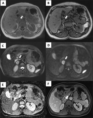
The aim of this retrospective study was to compare the MRI features between typical and atypical pheochromocytomas (Pheos) to specifically illustrate MRI features of atypical tumors for helping tumor diagnosis.
Methods
A total of 22 patients (14 women and 8 men, median age: 53 years, age range: 25–82 years) with Pheos evaluated using a 3 T MRI scanner were retrospectively collected; in particular, all patients had one tumor lesion, except in two cases who had two and three lesions, respectively, for a total of 25 tumor lesions.
Results
Of the total 25 tumor lesions included in our series, 12 lesions were classified as typical for their classical appearance on MRI (T1 hypointensity, T2 hyperintensity, no signal drop on T1 out-of-phase, restricted diffusion and persistent contrast enhancement). Conversely, the other 13 tumors were classified as having atypical lesions because they did not show the MRI features observed in typical Pheos; in particular, 3 lesions showed signal intensity suggestive of tumor hemorrhagic changes, 2 lesions were totally cystic with an internal fluid–fluid level and a thin capsula, 3 lesions showed predominantly cystic signal intensity with residual solid tissue in the peripheral capsula, and the remaining 5 lesions appeared as rounded partially cystic lesions with associated areas of solid tissue.
Conclusion
The imaging characterization of typical Pheos may be performed using MRI with specific imaging features; however, atypical Pheos represents a diagnostic challenge using MRI; in these tumors, cystic, necrotic, hemorrhagic, or fat changes may occur; thus, diagnostic pitfalls should be taken into consideration for MRI interpretation of such tumor type in clinical practice.
期刊介绍:
Clinical and Translational Imaging is an international journal that publishes timely, up-to-date summaries on clinical practice and translational research and clinical applications of approved and experimental radiopharmaceuticals for diagnostic and therapeutic purposes. Coverage includes such topics as advanced preclinical evidence in the fields of physics, dosimetry, radiation biology and radiopharmacy with relevance to applications in human subjects. The journal benefits a readership of nuclear medicine practitioners and allied professionals involved in molecular imaging and therapy.


 分享
分享
 求助内容:
求助内容: 应助结果提醒方式:
 扫码关注我们
