{"title":"Fluorescence Spectroscopy Based Identification of Pseudomonas Aeruginosa and Escherichia Coli Suspensions.","authors":"Sana Imtiaz, Muhammad Saleem","doi":"10.1007/s10895-024-03608-w","DOIUrl":null,"url":null,"abstract":"<p><p>In this article, Fluorescence spectroscopy has been employed for the identification of Pseudomonas aeruginosa (PA) and Escherichia coli (E. coli) in water suspension. Emission spectra of PA and E. coli suspensions have been acquired by using excitation wavelengths from 270 to 420 nm with steps of 10 nm to explore their spectral features. It has been found that the emission spectra of tryptophan, tyrosine, NADH and FAD, being the intracellular biomolecules present in both bacteria, can be used as fingerprints for their identification, differentiation and quantification. Both bacterial strains can clearly be differentiated from water and from each other by using λ<sub>ex</sub> 270-290 nm through spectral analysis and from λ<sub>ex</sub>: 300-500 nm by applying statistical analysis. Furthermore, calibration curves for different bacterial loads of PA and E. coli suspensions have been produced between colonies forming units per ml (CFUs/ml) the integrated intensities of their emission spectra. CFUs/ml of both bacterial suspensions have been determined through plate count method which was used as cross-reference for the analysis of emission spectra of both bacterial suspensions. These curves may be used to estimate CFU/ml of both PA and E. coli in unknown water suspensions by determining the integrating intensity of their emission spectra.</p>","PeriodicalId":15800,"journal":{"name":"Journal of Fluorescence","volume":" ","pages":"1381-1391"},"PeriodicalIF":3.1000,"publicationDate":"2025-03-01","publicationTypes":"Journal Article","fieldsOfStudy":null,"isOpenAccess":false,"openAccessPdf":"","citationCount":"0","resultStr":null,"platform":"Semanticscholar","paperid":null,"PeriodicalName":"Journal of Fluorescence","FirstCategoryId":"92","ListUrlMain":"https://doi.org/10.1007/s10895-024-03608-w","RegionNum":4,"RegionCategory":"化学","ArticlePicture":[],"TitleCN":null,"AbstractTextCN":null,"PMCID":null,"EPubDate":"2024/2/9 0:00:00","PubModel":"Epub","JCR":"Q2","JCRName":"BIOCHEMICAL RESEARCH METHODS","Score":null,"Total":0}
引用次数: 0
Abstract
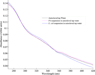
In this article, Fluorescence spectroscopy has been employed for the identification of Pseudomonas aeruginosa (PA) and Escherichia coli (E. coli) in water suspension. Emission spectra of PA and E. coli suspensions have been acquired by using excitation wavelengths from 270 to 420 nm with steps of 10 nm to explore their spectral features. It has been found that the emission spectra of tryptophan, tyrosine, NADH and FAD, being the intracellular biomolecules present in both bacteria, can be used as fingerprints for their identification, differentiation and quantification. Both bacterial strains can clearly be differentiated from water and from each other by using λex 270-290 nm through spectral analysis and from λex: 300-500 nm by applying statistical analysis. Furthermore, calibration curves for different bacterial loads of PA and E. coli suspensions have been produced between colonies forming units per ml (CFUs/ml) the integrated intensities of their emission spectra. CFUs/ml of both bacterial suspensions have been determined through plate count method which was used as cross-reference for the analysis of emission spectra of both bacterial suspensions. These curves may be used to estimate CFU/ml of both PA and E. coli in unknown water suspensions by determining the integrating intensity of their emission spectra.
期刊介绍:
Journal of Fluorescence is an international forum for the publication of peer-reviewed original articles that advance the practice of this established spectroscopic technique. Topics covered include advances in theory/and or data analysis, studies of the photophysics of aromatic molecules, solvent, and environmental effects, development of stationary or time-resolved measurements, advances in fluorescence microscopy, imaging, photobleaching/recovery measurements, and/or phosphorescence for studies of cell biology, chemical biology and the advanced uses of fluorescence in flow cytometry/analysis, immunology, high throughput screening/drug discovery, DNA sequencing/arrays, genomics and proteomics. Typical applications might include studies of macromolecular dynamics and conformation, intracellular chemistry, and gene expression. The journal also publishes papers that describe the synthesis and characterization of new fluorophores, particularly those displaying unique sensitivities and/or optical properties. In addition to original articles, the Journal also publishes reviews, rapid communications, short communications, letters to the editor, topical news articles, and technical and design notes.


 分享
分享
 求助内容:
求助内容: 应助结果提醒方式:
 扫码关注我们
