{"title":"Radiation evaluation assay using a human three-dimensional oral cancer model for clinical radiation therapy.","authors":"Lucie Sercombe , Kazuyo Igawa , Kenji Izumi","doi":"10.1016/j.talo.2024.100297","DOIUrl":null,"url":null,"abstract":"<div><p>With the development of various radiation-based cancer therapies, radiobiological evaluation methods instead of traditional clonogenic assays with monolayer single cell culture are required to bridge gaps in clinical data. Heterogeneity within cancer tissues is the reason for bridging the gap between basic and clinical research in cancer radiotherapy. To solve this problem, we investigated an evaluation assay using a three-dimensional (3D) model of cancer tissue. In this study, a 3D model consisting of tumor and stromal layers was used to compare and verify radiobiological effects with conventional two-dimensional (2D) methods. A significant difference in the response to radiation was observed between the 2D and 3D models. The relative number of cancer cells decreased with X-ray dose escalations in the 2D and 3D models. In contrast, the relative number of normal cells was quite different between the 2D and 3D models. Considering the ability of cells to recover from radiation-induced damage, the histological results of the 3D model were reflected in the clinical data. Histopathological analysis using a 3D model is a potential method for evaluating radiobiological effects on the tumor and tumor margins.</p></div>","PeriodicalId":436,"journal":{"name":"Talanta Open","volume":"9 ","pages":"Article 100297"},"PeriodicalIF":3.7000,"publicationDate":"2024-08-01","publicationTypes":"Journal Article","fieldsOfStudy":null,"isOpenAccess":false,"openAccessPdf":"https://www.sciencedirect.com/science/article/pii/S2666831924000110/pdfft?md5=bc5883f91ca2ca484638358c38d000fe&pid=1-s2.0-S2666831924000110-main.pdf","citationCount":"0","resultStr":null,"platform":"Semanticscholar","paperid":null,"PeriodicalName":"Talanta Open","FirstCategoryId":"1085","ListUrlMain":"https://www.sciencedirect.com/science/article/pii/S2666831924000110","RegionNum":0,"RegionCategory":null,"ArticlePicture":[],"TitleCN":null,"AbstractTextCN":null,"PMCID":null,"EPubDate":"2024/2/16 0:00:00","PubModel":"Epub","JCR":"Q1","JCRName":"CHEMISTRY, ANALYTICAL","Score":null,"Total":0}
引用次数: 0
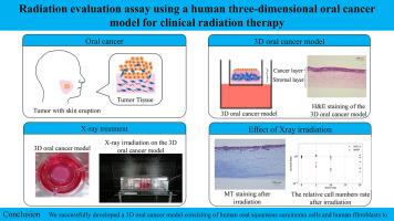
Abstract
With the development of various radiation-based cancer therapies, radiobiological evaluation methods instead of traditional clonogenic assays with monolayer single cell culture are required to bridge gaps in clinical data. Heterogeneity within cancer tissues is the reason for bridging the gap between basic and clinical research in cancer radiotherapy. To solve this problem, we investigated an evaluation assay using a three-dimensional (3D) model of cancer tissue. In this study, a 3D model consisting of tumor and stromal layers was used to compare and verify radiobiological effects with conventional two-dimensional (2D) methods. A significant difference in the response to radiation was observed between the 2D and 3D models. The relative number of cancer cells decreased with X-ray dose escalations in the 2D and 3D models. In contrast, the relative number of normal cells was quite different between the 2D and 3D models. Considering the ability of cells to recover from radiation-induced damage, the histological results of the 3D model were reflected in the clinical data. Histopathological analysis using a 3D model is a potential method for evaluating radiobiological effects on the tumor and tumor margins.
随着各种基于辐射的癌症疗法的发展,需要用放射生物学评估方法取代传统的单层单细胞培养克隆生成试验,以弥补临床数据的不足。癌症组织内部的异质性是弥合癌症放射治疗基础研究与临床研究之间差距的原因。为了解决这个问题,我们研究了一种使用癌症组织三维(3D)模型的评估检测方法。在这项研究中,我们使用由肿瘤层和基质层组成的三维模型来比较和验证传统二维(2D)方法的放射生物学效应。二维模型和三维模型对辐射的反应存在明显差异。在二维和三维模型中,癌细胞的相对数量随着 X 射线剂量的增加而减少。相比之下,二维和三维模型中正常细胞的相对数量则大不相同。考虑到细胞从辐射损伤中恢复的能力,三维模型的组织学结果反映在临床数据中。使用三维模型进行组织病理学分析是评估放射生物学对肿瘤和肿瘤边缘影响的一种潜在方法。


 分享
分享
 求助内容:
求助内容: 应助结果提醒方式:
 扫码关注我们
