Radiomics Features on Enhanced Computed Tomography Predict FOXP3 Expression and Clinical Prognosis in Patients with Head and Neck Squamous Cell Carcinoma.
Yi Wang, Juan Ye, Kai Zhou, Nian Chen, Gang Huang, Guangyong Feng, Guihai Zhang, Xiaoxia Gou
{"title":"Radiomics Features on Enhanced Computed Tomography Predict FOXP3 Expression and Clinical Prognosis in Patients with Head and Neck Squamous Cell Carcinoma.","authors":"Yi Wang, Juan Ye, Kai Zhou, Nian Chen, Gang Huang, Guangyong Feng, Guihai Zhang, Xiaoxia Gou","doi":"10.1007/s10278-023-00910-0","DOIUrl":null,"url":null,"abstract":"<p><p>Forkhead box P3 (FOXP3) has been identified as a novel molecular marker in various types of cancer. The present study assessed the expression of FOXP3 in patients with head and neck squamous cell carcinoma (HNSCC) and its potential as a clinical prognostic indicator, and developed a radiomics model based on enhanced computed tomography (CT) imaging. Data from 483 patients with HNSCC were downloaded from the Cancer Genome Atlas for FOXP3 prognostic analysis and enhanced CT images from 139 patients included in the Cancer Imaging Archives, which were subjected to the maximum relevance and minimum redundancy and recursive feature elimination algorithms for radiomics feature extraction and processing. Logistic regression was used to build a model for predicting FOXP3 expression. A prognostic scoring system for radiomics score (RS), FOXP3, and patient clinicopathological factors was established to predict patient survival. The area under the receiver operating characteristic (ROC) curve (AUC) and calibration curve and decision curve analysis (DCA) were used to evaluate model performance. Furthermore, the relationship between FOXP3 and the immune microenvironment, as well as the association between RS and immune checkpoint-related genes, was analyzed. Results of analysis revealed that patients with HNSCC and high FOXP3 mRNA expression exhibited better overall survival. Immune infiltration analysis revealed that FOXP3 had a positive correlation with CD4<sup> +</sup> and CD8<sup> +</sup> T cells and other immune cells. The 8 best radiomics features were selected to construct the radiomics model. In the FOXP3 expression prediction model, the AUC values were 0.707 and 0.702 for the training and validation sets, respectively. Additionally, the calibration curve and DCA demonstrated the positive diagnostic utility of the model. RS was correlated with immune checkpoint-related genes such as ICOS, CTLA4, and PDCD1. A predictive nomogram was established, the AUCs were 0.87, 0.787, and 0.801 at 12, 24, and 36 months, respectively, and DCA demonstrated the high clinical applicability of the nomogram. The enhanced CT radiomics model can predict expression of FOXP3 and prognosis in patients with HNSCC. As such, FOXP3 may be used as a novel prognostic marker to improve individualized clinical diagnosis and treatment decisions.</p>","PeriodicalId":516858,"journal":{"name":"Journal of imaging informatics in medicine","volume":" ","pages":"1323-1335"},"PeriodicalIF":0.0000,"publicationDate":"2024-08-01","publicationTypes":"Journal Article","fieldsOfStudy":null,"isOpenAccess":false,"openAccessPdf":"https://www.ncbi.nlm.nih.gov/pmc/articles/PMC11300763/pdf/","citationCount":"0","resultStr":null,"platform":"Semanticscholar","paperid":null,"PeriodicalName":"Journal of imaging informatics in medicine","FirstCategoryId":"1085","ListUrlMain":"https://doi.org/10.1007/s10278-023-00910-0","RegionNum":0,"RegionCategory":null,"ArticlePicture":[],"TitleCN":null,"AbstractTextCN":null,"PMCID":null,"EPubDate":"2024/2/20 0:00:00","PubModel":"Epub","JCR":"","JCRName":"","Score":null,"Total":0}
引用次数: 0
Abstract
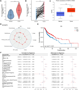
Forkhead box P3 (FOXP3) has been identified as a novel molecular marker in various types of cancer. The present study assessed the expression of FOXP3 in patients with head and neck squamous cell carcinoma (HNSCC) and its potential as a clinical prognostic indicator, and developed a radiomics model based on enhanced computed tomography (CT) imaging. Data from 483 patients with HNSCC were downloaded from the Cancer Genome Atlas for FOXP3 prognostic analysis and enhanced CT images from 139 patients included in the Cancer Imaging Archives, which were subjected to the maximum relevance and minimum redundancy and recursive feature elimination algorithms for radiomics feature extraction and processing. Logistic regression was used to build a model for predicting FOXP3 expression. A prognostic scoring system for radiomics score (RS), FOXP3, and patient clinicopathological factors was established to predict patient survival. The area under the receiver operating characteristic (ROC) curve (AUC) and calibration curve and decision curve analysis (DCA) were used to evaluate model performance. Furthermore, the relationship between FOXP3 and the immune microenvironment, as well as the association between RS and immune checkpoint-related genes, was analyzed. Results of analysis revealed that patients with HNSCC and high FOXP3 mRNA expression exhibited better overall survival. Immune infiltration analysis revealed that FOXP3 had a positive correlation with CD4 + and CD8 + T cells and other immune cells. The 8 best radiomics features were selected to construct the radiomics model. In the FOXP3 expression prediction model, the AUC values were 0.707 and 0.702 for the training and validation sets, respectively. Additionally, the calibration curve and DCA demonstrated the positive diagnostic utility of the model. RS was correlated with immune checkpoint-related genes such as ICOS, CTLA4, and PDCD1. A predictive nomogram was established, the AUCs were 0.87, 0.787, and 0.801 at 12, 24, and 36 months, respectively, and DCA demonstrated the high clinical applicability of the nomogram. The enhanced CT radiomics model can predict expression of FOXP3 and prognosis in patients with HNSCC. As such, FOXP3 may be used as a novel prognostic marker to improve individualized clinical diagnosis and treatment decisions.


 分享
分享

 求助内容:
求助内容: 应助结果提醒方式:
 扫码关注我们
