Thomas I. Mincherton, Stephanie J. Lam, Sarah E. Clarke, Henry Y. L. Hui, Jacques A. J. Malherbe, Hun S. Chuah, M. Hasib Sidiqi, Kathy A. Fuller, Wendy N. Erber
{"title":"Imaging flow cytometric detection of del(17p) in bone marrow and circulating plasma cells in multiple myeloma","authors":"Thomas I. Mincherton, Stephanie J. Lam, Sarah E. Clarke, Henry Y. L. Hui, Jacques A. J. Malherbe, Hun S. Chuah, M. Hasib Sidiqi, Kathy A. Fuller, Wendy N. Erber","doi":"10.1111/ijlh.14248","DOIUrl":null,"url":null,"abstract":"<div>\n \n \n <section>\n \n <h3> Background</h3>\n \n <p>Detection of del(17p) in myeloma is generally performed by fluorescence in situ hybridization (FISH) on a slide with analysis of up to 200 nuclei. The small cell sample analyzed makes this a low precision test. We report the utility of an automated FISH method, called “immuno-flowFISH”, to detect plasma cells with adverse prognostic risk del(17p) in bone marrow and blood samples of patients with myeloma.</p>\n </section>\n \n <section>\n \n <h3> Methods</h3>\n \n <p>Bone marrow (<i>n</i> = 31) and blood (<i>n</i> = 19) samples from 35 patients with myeloma were analyzed using immuno-flowFISH. Plasma cells were identified by CD38/CD138-immunophenotypic gating and assessed for the 17p locus and centromere of chromosome 17. Cells were acquired on an AMNIS ImageStreamX MkII imaging flow cytometer using INSPIRE software.</p>\n </section>\n \n <section>\n \n <h3> Results</h3>\n \n <p>Chromosome 17 abnormalities were identified in CD38/CD138-positive cells in bone marrow (6/31) and blood (4/19) samples when the percent plasma cell burden ranged from 0.03% to 100% of cells. Abnormalities could be identified in 14.5%–100% of plasma cells.</p>\n </section>\n \n <section>\n \n <h3> Conclusions</h3>\n \n <p>The “immuno-flowFISH” imaging flow cytometric method could detect del(17p) in plasma cells in both bone marrow and blood samples of myeloma patients. This method was also able to detect gains and losses of chromosome 17, which are also of prognostic significance. The lowest levels of 0.009% (bone marrow) and 0.001% (blood) for chromosome 17 abnormalities was below the detection limit of current FISH method. This method offers potential as a new means of identifying these prognostically important chromosomal defects, even when only rare cells are present and for serial disease monitoring.</p>\n </section>\n </div>","PeriodicalId":14120,"journal":{"name":"International Journal of Laboratory Hematology","volume":"46 3","pages":"495-502"},"PeriodicalIF":2.3000,"publicationDate":"2024-02-21","publicationTypes":"Journal Article","fieldsOfStudy":null,"isOpenAccess":false,"openAccessPdf":"https://onlinelibrary.wiley.com/doi/epdf/10.1111/ijlh.14248","citationCount":"0","resultStr":null,"platform":"Semanticscholar","paperid":null,"PeriodicalName":"International Journal of Laboratory Hematology","FirstCategoryId":"3","ListUrlMain":"https://onlinelibrary.wiley.com/doi/10.1111/ijlh.14248","RegionNum":4,"RegionCategory":"医学","ArticlePicture":[],"TitleCN":null,"AbstractTextCN":null,"PMCID":null,"EPubDate":"","PubModel":"","JCR":"Q3","JCRName":"HEMATOLOGY","Score":null,"Total":0}
引用次数: 0
Abstract
Background
Detection of del(17p) in myeloma is generally performed by fluorescence in situ hybridization (FISH) on a slide with analysis of up to 200 nuclei. The small cell sample analyzed makes this a low precision test. We report the utility of an automated FISH method, called “immuno-flowFISH”, to detect plasma cells with adverse prognostic risk del(17p) in bone marrow and blood samples of patients with myeloma.
Methods
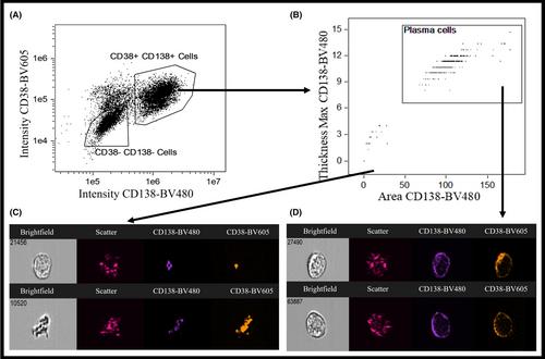
Bone marrow (n = 31) and blood (n = 19) samples from 35 patients with myeloma were analyzed using immuno-flowFISH. Plasma cells were identified by CD38/CD138-immunophenotypic gating and assessed for the 17p locus and centromere of chromosome 17. Cells were acquired on an AMNIS ImageStreamX MkII imaging flow cytometer using INSPIRE software.
Results
Chromosome 17 abnormalities were identified in CD38/CD138-positive cells in bone marrow (6/31) and blood (4/19) samples when the percent plasma cell burden ranged from 0.03% to 100% of cells. Abnormalities could be identified in 14.5%–100% of plasma cells.
Conclusions
The “immuno-flowFISH” imaging flow cytometric method could detect del(17p) in plasma cells in both bone marrow and blood samples of myeloma patients. This method was also able to detect gains and losses of chromosome 17, which are also of prognostic significance. The lowest levels of 0.009% (bone marrow) and 0.001% (blood) for chromosome 17 abnormalities was below the detection limit of current FISH method. This method offers potential as a new means of identifying these prognostically important chromosomal defects, even when only rare cells are present and for serial disease monitoring.
期刊介绍:
The International Journal of Laboratory Hematology provides a forum for the communication of new developments, research topics and the practice of laboratory haematology.
The journal publishes invited reviews, full length original articles, and correspondence.
The International Journal of Laboratory Hematology is the official journal of the International Society for Laboratory Hematology, which addresses the following sub-disciplines: cellular analysis, flow cytometry, haemostasis and thrombosis, molecular diagnostics, haematology informatics, haemoglobinopathies, point of care testing, standards and guidelines.
The journal was launched in 2006 as the successor to Clinical and Laboratory Hematology, which was first published in 1979. An active and positive editorial policy ensures that work of a high scientific standard is reported, in order to bridge the gap between practical and academic aspects of laboratory haematology.


 分享
分享
 求助内容:
求助内容: 应助结果提醒方式:
 扫码关注我们
