{"title":"Head-to-head comparison of [18F]FDG PET/MRI and [18F] FDG PET/CT for TNM staging in non-small cell lung cancer: a systematic review and meta-analysis","authors":"Zhiwei Li, Dianhan Sun, Anying Li, Yusheng Shu","doi":"10.1007/s40336-024-00622-7","DOIUrl":null,"url":null,"abstract":"<h3 data-test=\"abstract-sub-heading\">Purpose</h3><p>This study aimed to compare the diagnostic accuracy of [<sup>18</sup>F]fluorodeoxyglucose (FDG) positron emission tomography (PET)/ magnetic resonance imaging (MRI) and [<sup>18</sup>F]FDG PET/ computed tomography (CT) in tumor–node–metastasis staging of non-small-cell lung cancer.</p><h3 data-test=\"abstract-sub-heading\">Methods</h3><p>The study followed the Preferred Reporting Items for Systematic Reviews and Meta-Analyses for Diagnostic Test Accuracy (PRISMA-DTA) guidelines and retrieved all accessible studies from the Embase, PubMed, and Web of Science databases up to December 2022. Only studies in which both [<sup>18</sup>F]FDG PET/MRI and [<sup>18</sup>F]FDG PET/CT were conducted on each individual patient were included. Two researchers independently extracted data on study characteristics and assessed the methodological quality using the Quality Assessment of Diagnostic Accuracy Studies (QUADAS-2) tool.</p><h3 data-test=\"abstract-sub-heading\">Results</h3><p>A total of 539 patients in eight studies were included in this analysis. For T staging, the pooled sensitivity of [<sup>18</sup>F]FDG PET/CT was 0.90 (95% confidence interval [CI]: 0.81–0.96) and specificity of 0.97 (95% CI: 0.89–1.00), with corresponding values for [<sup>18</sup>F]FDG PET/MRI of 0.88 (95% CI: 0.78–0.94) and 0.95 (95% CI: 0.87–0.99), respectively. For N staging, the pooled sensitivity of [<sup>18</sup>F] FDG PET/CT was 0.70 (95% CI: 0.63–0.76), the specificity of 0.92 (95% CI: 0.88–0.95), and the area under the curve (AUC) was 0.90 (standard error [SE] = 0.06). The corresponding values for [<sup>18</sup>F]FDG PET/MRI were 0.71 (95% CI: 0.65–0.77), 0.91 (95% CI: 0.87–0.94) and 0.88 (SE = 0.06), respectively. For M staging, the pooled sensitivity was 0.79 (95% CI: 0.62–0.91), the specificity was 0.94 (95% CI: 0.90–0.97), and AUC was 0.96 (SE = 0.03) for [<sup>18</sup>F]FDG PET/CT. The corresponding values were 0.82 (95% CI: 0.70–0.91), 0.96 (95% CI: 0.93–0.98), and 0.94 (SE = 0.03), respectively, for [<sup>18</sup>F]FDG PET/MRI.</p><h3 data-test=\"abstract-sub-heading\">Conclusions</h3><p>According to the pooled data, [<sup>18</sup>F]FDG PET/CT performed slightly better in terms of T staging than [<sup>18</sup>F]FDG PET/MRI. In contrast, with regard to N staging and M staging the staging accuracy of both imaging techniques was comparable. To ensure that results are reliable, more high-level investigations will be required to assess these imaging modalities, in addition to optimized PET/MRI procedures.</p>","PeriodicalId":48600,"journal":{"name":"Clinical and Translational Imaging","volume":"2015 1","pages":""},"PeriodicalIF":1.6000,"publicationDate":"2024-02-23","publicationTypes":"Journal Article","fieldsOfStudy":null,"isOpenAccess":false,"openAccessPdf":"","citationCount":"0","resultStr":null,"platform":"Semanticscholar","paperid":null,"PeriodicalName":"Clinical and Translational Imaging","FirstCategoryId":"3","ListUrlMain":"https://doi.org/10.1007/s40336-024-00622-7","RegionNum":4,"RegionCategory":"医学","ArticlePicture":[],"TitleCN":null,"AbstractTextCN":null,"PMCID":null,"EPubDate":"","PubModel":"","JCR":"Q2","JCRName":"RADIOLOGY, NUCLEAR MEDICINE & MEDICAL IMAGING","Score":null,"Total":0}
引用次数: 0
Abstract
Purpose
This study aimed to compare the diagnostic accuracy of [18F]fluorodeoxyglucose (FDG) positron emission tomography (PET)/ magnetic resonance imaging (MRI) and [18F]FDG PET/ computed tomography (CT) in tumor–node–metastasis staging of non-small-cell lung cancer.
Methods
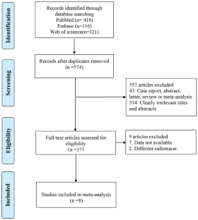
The study followed the Preferred Reporting Items for Systematic Reviews and Meta-Analyses for Diagnostic Test Accuracy (PRISMA-DTA) guidelines and retrieved all accessible studies from the Embase, PubMed, and Web of Science databases up to December 2022. Only studies in which both [18F]FDG PET/MRI and [18F]FDG PET/CT were conducted on each individual patient were included. Two researchers independently extracted data on study characteristics and assessed the methodological quality using the Quality Assessment of Diagnostic Accuracy Studies (QUADAS-2) tool.
Results
A total of 539 patients in eight studies were included in this analysis. For T staging, the pooled sensitivity of [18F]FDG PET/CT was 0.90 (95% confidence interval [CI]: 0.81–0.96) and specificity of 0.97 (95% CI: 0.89–1.00), with corresponding values for [18F]FDG PET/MRI of 0.88 (95% CI: 0.78–0.94) and 0.95 (95% CI: 0.87–0.99), respectively. For N staging, the pooled sensitivity of [18F] FDG PET/CT was 0.70 (95% CI: 0.63–0.76), the specificity of 0.92 (95% CI: 0.88–0.95), and the area under the curve (AUC) was 0.90 (standard error [SE] = 0.06). The corresponding values for [18F]FDG PET/MRI were 0.71 (95% CI: 0.65–0.77), 0.91 (95% CI: 0.87–0.94) and 0.88 (SE = 0.06), respectively. For M staging, the pooled sensitivity was 0.79 (95% CI: 0.62–0.91), the specificity was 0.94 (95% CI: 0.90–0.97), and AUC was 0.96 (SE = 0.03) for [18F]FDG PET/CT. The corresponding values were 0.82 (95% CI: 0.70–0.91), 0.96 (95% CI: 0.93–0.98), and 0.94 (SE = 0.03), respectively, for [18F]FDG PET/MRI.
Conclusions
According to the pooled data, [18F]FDG PET/CT performed slightly better in terms of T staging than [18F]FDG PET/MRI. In contrast, with regard to N staging and M staging the staging accuracy of both imaging techniques was comparable. To ensure that results are reliable, more high-level investigations will be required to assess these imaging modalities, in addition to optimized PET/MRI procedures.
期刊介绍:
Clinical and Translational Imaging is an international journal that publishes timely, up-to-date summaries on clinical practice and translational research and clinical applications of approved and experimental radiopharmaceuticals for diagnostic and therapeutic purposes. Coverage includes such topics as advanced preclinical evidence in the fields of physics, dosimetry, radiation biology and radiopharmacy with relevance to applications in human subjects. The journal benefits a readership of nuclear medicine practitioners and allied professionals involved in molecular imaging and therapy.


 分享
分享
 求助内容:
求助内容: 应助结果提醒方式:
 扫码关注我们
