{"title":"Revision of the muscular system in the brachiopod Novocrania anomala using 3D reconstruction: Functional and paleontological significance","authors":"Feodor A. Plandin, Elena N. Temereva","doi":"10.1002/jmor.21685","DOIUrl":null,"url":null,"abstract":"<p>The musculature is one of the best studied organ systems in brachiopods, being approachable not only by dissecting recent species of brachiopods, but also by exploring muscle scars in fossil material. In the present study, the muscular anatomy of <i>Novocrania anomala</i> is studied using 3D reconstructions based on microcomputed tomography. Muscles of <i>N. anomala</i> may be subdivided into two groups: those related to movements of the lophophore, and those connected to movements of shell valves. Muscles, their morphology and possible functions, such as brachial protractors, elevators, and retractors, as well as anterior adductors, are described and discussed. We also provide the discussion of craniid muscle terminology, consider the valve-opening mechanism. The investigation of muscle scars on dorsal valves supports the conclusion that the shape of muscle scars should be used for description and distinction of recent and extinct species only when visible distinctness cannot be explained by substrate differences. This study, which is aimed at improving our understanding the anatomy and functioning of muscles in craniids, will be useful not only for zoologists, but also for paleontologists.</p>","PeriodicalId":16528,"journal":{"name":"Journal of Morphology","volume":"285 3","pages":""},"PeriodicalIF":1.4000,"publicationDate":"2024-03-11","publicationTypes":"Journal Article","fieldsOfStudy":null,"isOpenAccess":false,"openAccessPdf":"","citationCount":"0","resultStr":null,"platform":"Semanticscholar","paperid":null,"PeriodicalName":"Journal of Morphology","FirstCategoryId":"3","ListUrlMain":"https://onlinelibrary.wiley.com/doi/10.1002/jmor.21685","RegionNum":4,"RegionCategory":"医学","ArticlePicture":[],"TitleCN":null,"AbstractTextCN":null,"PMCID":null,"EPubDate":"","PubModel":"","JCR":"Q2","JCRName":"ANATOMY & MORPHOLOGY","Score":null,"Total":0}
引用次数: 0
Abstract
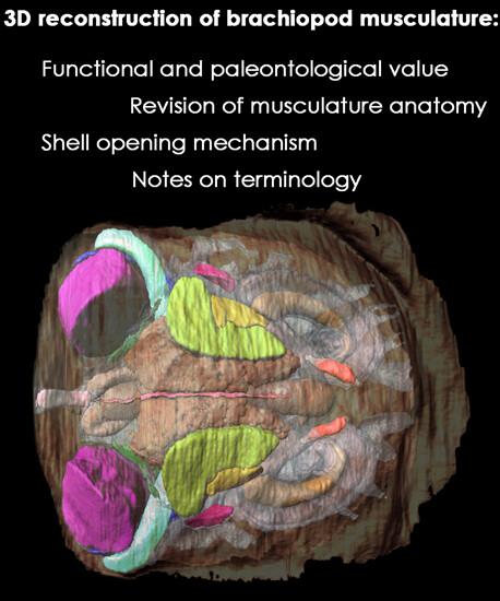
The musculature is one of the best studied organ systems in brachiopods, being approachable not only by dissecting recent species of brachiopods, but also by exploring muscle scars in fossil material. In the present study, the muscular anatomy of Novocrania anomala is studied using 3D reconstructions based on microcomputed tomography. Muscles of N. anomala may be subdivided into two groups: those related to movements of the lophophore, and those connected to movements of shell valves. Muscles, their morphology and possible functions, such as brachial protractors, elevators, and retractors, as well as anterior adductors, are described and discussed. We also provide the discussion of craniid muscle terminology, consider the valve-opening mechanism. The investigation of muscle scars on dorsal valves supports the conclusion that the shape of muscle scars should be used for description and distinction of recent and extinct species only when visible distinctness cannot be explained by substrate differences. This study, which is aimed at improving our understanding the anatomy and functioning of muscles in craniids, will be useful not only for zoologists, but also for paleontologists.
期刊介绍:
The Journal of Morphology welcomes articles of original research in cytology, protozoology, embryology, and general morphology. Articles generally should not exceed 35 printed pages. Preliminary notices or articles of a purely descriptive morphological or taxonomic nature are not included. No paper which has already been published will be accepted, nor will simultaneous publications elsewhere be allowed.
The Journal of Morphology publishes research in functional, comparative, evolutionary and developmental morphology from vertebrates and invertebrates. Human and veterinary anatomy or paleontology are considered when an explicit connection to neontological animal morphology is presented, and the paper contains relevant information for the community of animal morphologists. Based on our long tradition, we continue to seek publishing the best papers in animal morphology.


 分享
分享
 求助内容:
求助内容: 应助结果提醒方式:
 扫码关注我们
