Shihao Yang, Meng Jiao, Jing Xiang, Neel Fotedar, Hai Sun, Feng Liu
{"title":"Rejuvenating classical brain electrophysiology source localization methods with spatial graph Fourier filters for source extents estimation.","authors":"Shihao Yang, Meng Jiao, Jing Xiang, Neel Fotedar, Hai Sun, Feng Liu","doi":"10.1186/s40708-024-00221-2","DOIUrl":null,"url":null,"abstract":"<p><p>EEG/MEG source imaging (ESI) aims to find the underlying brain sources to explain the observed EEG or MEG measurement. Multiple classical approaches have been proposed to solve the ESI problem based on different neurophysiological assumptions. To support clinical decision-making, it is important to estimate not only the exact location of the source signal but also the extended source activation regions. Existing methods may render over-diffuse or sparse solutions, which limit the source extent estimation accuracy. In this work, we leverage the graph structures defined in the 3D mesh of the brain and the spatial graph Fourier transform (GFT) to decompose the spatial graph structure into sub-spaces of low-, medium-, and high-frequency basis. We propose to use the low-frequency basis of spatial graph filters to approximate the extended areas of brain activation and embed the GFT into the classical ESI methods. We validated the classical source localization methods with the corresponding improved version using GFT in both synthetic data and real data. We found the proposed method can effectively reconstruct focal source patterns and significantly improve the performance compared to the classical algorithms.</p>","PeriodicalId":37465,"journal":{"name":"Brain Informatics","volume":"11 1","pages":"8"},"PeriodicalIF":4.5000,"publicationDate":"2024-03-12","publicationTypes":"Journal Article","fieldsOfStudy":null,"isOpenAccess":false,"openAccessPdf":"https://www.ncbi.nlm.nih.gov/pmc/articles/PMC10933195/pdf/","citationCount":"0","resultStr":null,"platform":"Semanticscholar","paperid":null,"PeriodicalName":"Brain Informatics","FirstCategoryId":"1085","ListUrlMain":"https://doi.org/10.1186/s40708-024-00221-2","RegionNum":0,"RegionCategory":null,"ArticlePicture":[],"TitleCN":null,"AbstractTextCN":null,"PMCID":null,"EPubDate":"","PubModel":"","JCR":"Q1","JCRName":"Computer Science","Score":null,"Total":0}
引用次数: 0
Abstract
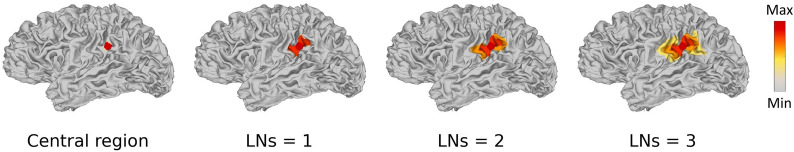
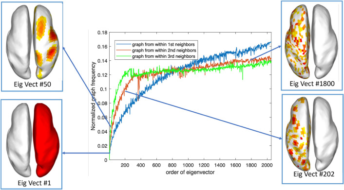
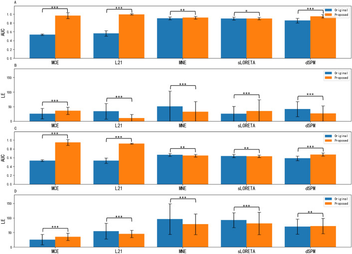
EEG/MEG source imaging (ESI) aims to find the underlying brain sources to explain the observed EEG or MEG measurement. Multiple classical approaches have been proposed to solve the ESI problem based on different neurophysiological assumptions. To support clinical decision-making, it is important to estimate not only the exact location of the source signal but also the extended source activation regions. Existing methods may render over-diffuse or sparse solutions, which limit the source extent estimation accuracy. In this work, we leverage the graph structures defined in the 3D mesh of the brain and the spatial graph Fourier transform (GFT) to decompose the spatial graph structure into sub-spaces of low-, medium-, and high-frequency basis. We propose to use the low-frequency basis of spatial graph filters to approximate the extended areas of brain activation and embed the GFT into the classical ESI methods. We validated the classical source localization methods with the corresponding improved version using GFT in both synthetic data and real data. We found the proposed method can effectively reconstruct focal source patterns and significantly improve the performance compared to the classical algorithms.
EEG/MEG 信号源成像(ESI)旨在找到潜在的大脑信号源,以解释观察到的 EEG 或 MEG 测量结果。基于不同的神经生理学假设,人们提出了多种经典方法来解决 ESI 问题。为了支持临床决策,重要的是不仅要估计源信号的确切位置,还要估计扩展的源激活区域。现有方法可能会产生过度弥散或稀疏的解决方案,从而限制了源范围估计的准确性。在这项工作中,我们利用大脑三维网格中定义的图结构和空间图傅里叶变换(GFT),将空间图结构分解为低频、中频和高频基础子空间。我们建议使用空间图滤波器的低频基础来近似大脑激活的扩展区域,并将 GFT 嵌入到经典的 ESI 方法中。我们在合成数据和真实数据中验证了经典源定位方法和使用 GFT 的相应改进版本。我们发现,与经典算法相比,所提出的方法能有效重建焦点源模式并显著提高性能。
期刊介绍:
Brain Informatics is an international, peer-reviewed, interdisciplinary open-access journal published under the brand SpringerOpen, which provides a unique platform for researchers and practitioners to disseminate original research on computational and informatics technologies related to brain. This journal addresses the computational, cognitive, physiological, biological, physical, ecological and social perspectives of brain informatics. It also welcomes emerging information technologies and advanced neuro-imaging technologies, such as big data analytics and interactive knowledge discovery related to various large-scale brain studies and their applications. This journal will publish high-quality original research papers, brief reports and critical reviews in all theoretical, technological, clinical and interdisciplinary studies that make up the field of brain informatics and its applications in brain-machine intelligence, brain-inspired intelligent systems, mental health and brain disorders, etc. The scope of papers includes the following five tracks: Track 1: Cognitive and Computational Foundations of Brain Science Track 2: Human Information Processing Systems Track 3: Brain Big Data Analytics, Curation and Management Track 4: Informatics Paradigms for Brain and Mental Health Research Track 5: Brain-Machine Intelligence and Brain-Inspired Computing


 分享
分享
 求助内容:
求助内容: 应助结果提醒方式:
 扫码关注我们
