Yangkang Zheng , Pengyu Wang , Li Zhao , Lianping Xing , Hao Xu , Ning Li , Yongjian Zhao , Qi Shi , Qianqian Liang , YongJun Wang
{"title":"A novel therapy for fracture healing by increasing lymphatic drainage","authors":"Yangkang Zheng , Pengyu Wang , Li Zhao , Lianping Xing , Hao Xu , Ning Li , Yongjian Zhao , Qi Shi , Qianqian Liang , YongJun Wang","doi":"10.1016/j.jot.2024.02.001","DOIUrl":null,"url":null,"abstract":"<div><h3>Background</h3><p>The musculoskeletal system contains an extensive network of lymphatic vessels. Decreased lymph flow of the draining collecting lymphatics usually occurs in clinic after traumatic fractures. However, whether defects in lymphatic drainage can affect fracture healing is unclear.</p></div><div><h3>Methods</h3><p>To investigate the effect of lymphatic dysfunction on fracture healing, we used a selective VEGFR3 tyrosine kinase inhibitor to treat tibial fractured mice for 5 weeks versus a vehicle-treated control. To ensure successfully establishing deceased lymphatic drainage model for fractured mice, we measured lymphatic clearance by near infrared indocyanine green lymphatic imaging (NIR-ICG) and the volume of the draining popliteal lymph nodes (PLNs) by ultrasound at the whole phases of fracture healing. In addition, hindlimb edema from day 0 to day 7 post-fracture, pain sensation by Hargreaves test at day 1 post-fracture, bone histomorphometry by micro-CT and callus composition by Alcian Blue-Hematoxylin/Orange G staining at day 14 post-fracture, and bone quality by biomechanical testing at day 35 post-fracture were applied to evaluate fracture healing. To promote fracture healing via increasing lymphatic drainage, we then treated fractured mice with anti-mouse podoplanin (PDPN) neutralizing antibody or isotype IgG antibody for 1 week to observe lymphatic drainage function and assess bone repair as methods described above.</p></div><div><h3>Results</h3><p>Compared to vehicle-treated group, SAR-treatment group significantly decreased lymphatic clearance and the volume of draining PLNs. SAR-treatment group significantly increased soft tissue swelling, and reduced bone volume (BV)/tissue volume (TV), trabecular number (Tb.N), woven bone and biomechanical properties of fracture callus. In addition, anti-PDPN treated group significantly reduced the number of CD41<sup>+</sup> platelets in PLNs and increased the number of pulsatile lymphatic vessels, lymphatic clearance and the volume of PLNs. Moreover, anti-PDPN treated group significantly reduced hindlimb edema and pain sensation and increased BV/TV, trabecular number (Tb.Th), woven bone and biomechanical properties of fracture callus.</p></div><div><h3>Conclusions</h3><p>Inhibition of proper lymphatic drainage function delayed fracture healing. Use of a anti-PDPN neutralizing antibody reduced lymphatic platelet thrombosis (LPT), increased lymphatic drainage and improved fracture healing.</p></div><div><h3>The translational potential of this article</h3><p>(1) We demonstrated lymphatic drainage function is crucial for fracture healing. (2) To unblock the lymphatic drainage and prevent the risk of bleeding and mortality by blood thinner, we demonstrated PDPN neutralizing antibody is a novel and safe way forward in the treatment of bone fracture healing by eliminating LPT and increasing lymphatic drainage.</p></div>","PeriodicalId":16636,"journal":{"name":"Journal of Orthopaedic Translation","volume":"45 ","pages":"Pages 66-74"},"PeriodicalIF":5.9000,"publicationDate":"2024-03-01","publicationTypes":"Journal Article","fieldsOfStudy":null,"isOpenAccess":false,"openAccessPdf":"https://www.sciencedirect.com/science/article/pii/S2214031X24000147/pdfft?md5=42170eb64192690edc2f138fdd8bc4fd&pid=1-s2.0-S2214031X24000147-main.pdf","citationCount":"0","resultStr":null,"platform":"Semanticscholar","paperid":null,"PeriodicalName":"Journal of Orthopaedic Translation","FirstCategoryId":"3","ListUrlMain":"https://www.sciencedirect.com/science/article/pii/S2214031X24000147","RegionNum":1,"RegionCategory":"医学","ArticlePicture":[],"TitleCN":null,"AbstractTextCN":null,"PMCID":null,"EPubDate":"2024/3/13 0:00:00","PubModel":"Epub","JCR":"Q1","JCRName":"ORTHOPEDICS","Score":null,"Total":0}
引用次数: 0
Abstract
Background
The musculoskeletal system contains an extensive network of lymphatic vessels. Decreased lymph flow of the draining collecting lymphatics usually occurs in clinic after traumatic fractures. However, whether defects in lymphatic drainage can affect fracture healing is unclear.
Methods
To investigate the effect of lymphatic dysfunction on fracture healing, we used a selective VEGFR3 tyrosine kinase inhibitor to treat tibial fractured mice for 5 weeks versus a vehicle-treated control. To ensure successfully establishing deceased lymphatic drainage model for fractured mice, we measured lymphatic clearance by near infrared indocyanine green lymphatic imaging (NIR-ICG) and the volume of the draining popliteal lymph nodes (PLNs) by ultrasound at the whole phases of fracture healing. In addition, hindlimb edema from day 0 to day 7 post-fracture, pain sensation by Hargreaves test at day 1 post-fracture, bone histomorphometry by micro-CT and callus composition by Alcian Blue-Hematoxylin/Orange G staining at day 14 post-fracture, and bone quality by biomechanical testing at day 35 post-fracture were applied to evaluate fracture healing. To promote fracture healing via increasing lymphatic drainage, we then treated fractured mice with anti-mouse podoplanin (PDPN) neutralizing antibody or isotype IgG antibody for 1 week to observe lymphatic drainage function and assess bone repair as methods described above.
Results
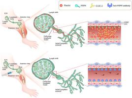
Compared to vehicle-treated group, SAR-treatment group significantly decreased lymphatic clearance and the volume of draining PLNs. SAR-treatment group significantly increased soft tissue swelling, and reduced bone volume (BV)/tissue volume (TV), trabecular number (Tb.N), woven bone and biomechanical properties of fracture callus. In addition, anti-PDPN treated group significantly reduced the number of CD41+ platelets in PLNs and increased the number of pulsatile lymphatic vessels, lymphatic clearance and the volume of PLNs. Moreover, anti-PDPN treated group significantly reduced hindlimb edema and pain sensation and increased BV/TV, trabecular number (Tb.Th), woven bone and biomechanical properties of fracture callus.
Conclusions
Inhibition of proper lymphatic drainage function delayed fracture healing. Use of a anti-PDPN neutralizing antibody reduced lymphatic platelet thrombosis (LPT), increased lymphatic drainage and improved fracture healing.
The translational potential of this article
(1) We demonstrated lymphatic drainage function is crucial for fracture healing. (2) To unblock the lymphatic drainage and prevent the risk of bleeding and mortality by blood thinner, we demonstrated PDPN neutralizing antibody is a novel and safe way forward in the treatment of bone fracture healing by eliminating LPT and increasing lymphatic drainage.
期刊介绍:
The Journal of Orthopaedic Translation (JOT) is the official peer-reviewed, open access journal of the Chinese Speaking Orthopaedic Society (CSOS) and the International Chinese Musculoskeletal Research Society (ICMRS). It is published quarterly, in January, April, July and October, by Elsevier.


 分享
分享
 求助内容:
求助内容: 应助结果提醒方式:
 扫码关注我们
