{"title":"Automatic estimation of hallux valgus angle using deep neural network with axis-based annotation.","authors":"Ryutaro Takeda, Hiroyasu Mizuhara, Akihiro Uchio, Toshiko Iidaka, Kenta Makabe, Taro Kasai, Yasunori Omata, Noriko Yoshimura, Sakae Tanaka, Takumi Matsumoto","doi":"10.1007/s00256-024-04618-2","DOIUrl":null,"url":null,"abstract":"<p><strong>Objectives: </strong>We developed the deep neural network (DNN) model to automatically measure hallux valgus angle (HVA) and intermetatarsal angle (IMA) on foot radiographs. The objective is to assess the accuracy of the model by comparing to the manual measurement of foot and ankle surgeons.</p><p><strong>Materials and methods: </strong>A DNN was developed to predict the bone axes of the first proximal phalanx and all metatarsals from the first to the fifth in foot radiographs. The dataset used for model development consisted of 1798 radiographs collected from a population-based cohort and patients at our foot and ankle clinic. The retrospective validation cohort comprised of 92 radiographs obtained from 92 consecutive patients visiting our foot and ankle clinic. The mean absolute error (MAE) between automatic measurements by the model and the median of manual measurements by three foot and ankle surgeons was compared to 3° using one-tailed t-test and was also compared to the inter-rater difference in manual measurements among the three surgeons using two-tailed paired t-test.</p><p><strong>Results: </strong>The MAE for HVA was 1.3° (upper limit of 95% CI 1.6°), and this was significantly smaller than the inter-rater difference of 2.0 ± 0.2° among the surgeons, demonstrating the superior accuracy of the model. In contrast, the MAE for IMA was 0.8° (upper limit of 95% CI 1.0°) that showed no significant difference from the inter-rater difference of 1.0 ± 0.1° among the surgeons.</p><p><strong>Conclusion: </strong>Our model demonstrated the ability to measure the HVA and IMA with an accuracy comparable to that of specialists.</p>","PeriodicalId":21783,"journal":{"name":"Skeletal Radiology","volume":" ","pages":"2357-2366"},"PeriodicalIF":2.2000,"publicationDate":"2024-11-01","publicationTypes":"Journal Article","fieldsOfStudy":null,"isOpenAccess":false,"openAccessPdf":"https://www.ncbi.nlm.nih.gov/pmc/articles/PMC11410836/pdf/","citationCount":"0","resultStr":null,"platform":"Semanticscholar","paperid":null,"PeriodicalName":"Skeletal Radiology","FirstCategoryId":"3","ListUrlMain":"https://doi.org/10.1007/s00256-024-04618-2","RegionNum":3,"RegionCategory":"医学","ArticlePicture":[],"TitleCN":null,"AbstractTextCN":null,"PMCID":null,"EPubDate":"2024/3/13 0:00:00","PubModel":"Epub","JCR":"Q2","JCRName":"ORTHOPEDICS","Score":null,"Total":0}
引用次数: 0
Abstract
Objectives: We developed the deep neural network (DNN) model to automatically measure hallux valgus angle (HVA) and intermetatarsal angle (IMA) on foot radiographs. The objective is to assess the accuracy of the model by comparing to the manual measurement of foot and ankle surgeons.
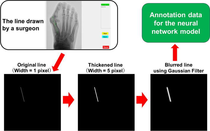
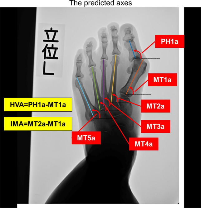
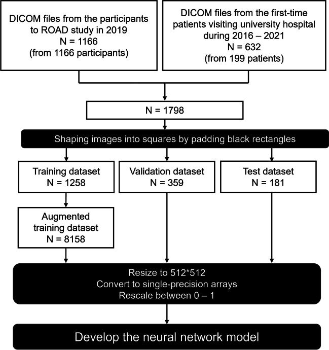
Materials and methods: A DNN was developed to predict the bone axes of the first proximal phalanx and all metatarsals from the first to the fifth in foot radiographs. The dataset used for model development consisted of 1798 radiographs collected from a population-based cohort and patients at our foot and ankle clinic. The retrospective validation cohort comprised of 92 radiographs obtained from 92 consecutive patients visiting our foot and ankle clinic. The mean absolute error (MAE) between automatic measurements by the model and the median of manual measurements by three foot and ankle surgeons was compared to 3° using one-tailed t-test and was also compared to the inter-rater difference in manual measurements among the three surgeons using two-tailed paired t-test.
Results: The MAE for HVA was 1.3° (upper limit of 95% CI 1.6°), and this was significantly smaller than the inter-rater difference of 2.0 ± 0.2° among the surgeons, demonstrating the superior accuracy of the model. In contrast, the MAE for IMA was 0.8° (upper limit of 95% CI 1.0°) that showed no significant difference from the inter-rater difference of 1.0 ± 0.1° among the surgeons.
Conclusion: Our model demonstrated the ability to measure the HVA and IMA with an accuracy comparable to that of specialists.
目的:我们开发了深度神经网络(DNN)模型,用于自动测量足部X光片上的外翻角(HVA)和跖间角(IMA)。目的是通过与足踝外科医生的人工测量进行比较,评估该模型的准确性:开发了 DNN 来预测足部 X 光片中第一近节指骨和第一至第五节所有跖骨的骨轴。用于模型开发的数据集包括从人群队列和足踝诊所患者处收集的 1798 张射线照片。回顾性验证队列包括从 92 名连续到我们足踝诊所就诊的患者处获得的 92 张射线照片。使用单尾 t 检验比较了模型自动测量值与三位足踝外科医生手工测量值中位数之间的平均绝对误差(MAE)与 3°之间的差异,并使用双尾配对 t 检验比较了三位外科医生手工测量值之间的差异:结果:HVA 的 MAE 为 1.3°(95% CI 上限为 1.6°),明显小于外科医生之间 2.0 ± 0.2°的评分者间差异,这表明该模型具有更高的准确性。相比之下,IMA的MAE为0.8°(95% CI上限为1.0°),与外科医生之间1.0 ± 0.1°的评分差异无明显差异:我们的模型证明了测量 HVA 和 IMA 的能力,其准确性可与专科医生媲美。
期刊介绍:
Skeletal Radiology provides a forum for the dissemination of current knowledge and information dealing with disorders of the musculoskeletal system including the spine. While emphasizing the radiological aspects of the many varied skeletal abnormalities, the journal also adopts an interdisciplinary approach, reflecting the membership of the International Skeletal Society. Thus, the anatomical, pathological, physiological, clinical, metabolic and epidemiological aspects of the many entities affecting the skeleton receive appropriate consideration.
This is the Journal of the International Skeletal Society and the Official Journal of the Society of Skeletal Radiology and the Australasian Musculoskelelal Imaging Group.


 分享
分享
 求助内容:
求助内容: 应助结果提醒方式:
 扫码关注我们
