Riccardo Cristiani, Fabian van de Bunt, Joanna Kvist, Anders Stålman
{"title":"High prevalence of associated injuries in anterior cruciate ligament tears: A detailed magnetic resonance imaging analysis of 254 patients.","authors":"Riccardo Cristiani, Fabian van de Bunt, Joanna Kvist, Anders Stålman","doi":"10.1007/s00256-024-04665-9","DOIUrl":null,"url":null,"abstract":"<p><strong>Objectives: </strong>To evaluate the type and prevalence of associated injuries by using magnetic resonance imaging (MRI) in patients with anterior cruciate ligament (ACL) tears.</p><p><strong>Methods: </strong>Data from the Natural Corollaries and Recovery after ACL injury multicenter longitudinal cohort study were analyzed. Between May 2016 and October 2018, patients aged between 15 and 40 years, who had experienced an ACL tear within the last 6 weeks and sought medical attention at one of seven healthcare clinics in Sweden, were invited to participate. The mean time from injury to MRI was 19.6 ± 15.2 days. An orthopedic knee surgeon and a musculoskeletal radiologist reviewed all the MRI scans. The following structures were assessed: posterior cruciate ligament (PCL), medial collateral ligament (MCL) complex, lateral collateral ligament (LCL), popliteus tendon, medial meniscus (MM), lateral meniscus (LM), and cartilage. In addition, the presence of bone bruising, impaction fractures in the lateral femoral condyle (LFC) or posterolateral tibia (PLT), and Segond fractures were also assessed. RESULTS: A total of 254 patients (48.4% males) with a mean age of 25.4 ± 7.1 years were included. The prevalence of associated injuries was as follows: PCL (0.4%), MCL {41.3% [superficial MCL and deep MCL (dMCL) 16.5%; isolated dMCL 24.8%]}, LCL (2.4%), MM (57.4%), LM (25.2%), cartilage (15.0%), bone bruising (92.9%), impaction fracture in the LFC (45.7%) and PLT (4.7%), and Segond fracture (7.5%).</p><p><strong>Conclusions: </strong>The prevalence of associated injuries in patients with ACL tears was high. The findings reported in this study may serve as a reference tool for orthopedic surgeons and radiologists in the diagnosis of associated injuries using MRI in patients with ACL tears.</p>","PeriodicalId":21783,"journal":{"name":"Skeletal Radiology","volume":" ","pages":"2417-2427"},"PeriodicalIF":2.2000,"publicationDate":"2024-11-01","publicationTypes":"Journal Article","fieldsOfStudy":null,"isOpenAccess":false,"openAccessPdf":"https://www.ncbi.nlm.nih.gov/pmc/articles/PMC11410909/pdf/","citationCount":"0","resultStr":null,"platform":"Semanticscholar","paperid":null,"PeriodicalName":"Skeletal Radiology","FirstCategoryId":"3","ListUrlMain":"https://doi.org/10.1007/s00256-024-04665-9","RegionNum":3,"RegionCategory":"医学","ArticlePicture":[],"TitleCN":null,"AbstractTextCN":null,"PMCID":null,"EPubDate":"2024/3/27 0:00:00","PubModel":"Epub","JCR":"Q2","JCRName":"ORTHOPEDICS","Score":null,"Total":0}
引用次数: 0
Abstract
Objectives: To evaluate the type and prevalence of associated injuries by using magnetic resonance imaging (MRI) in patients with anterior cruciate ligament (ACL) tears.
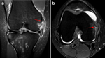
Methods: Data from the Natural Corollaries and Recovery after ACL injury multicenter longitudinal cohort study were analyzed. Between May 2016 and October 2018, patients aged between 15 and 40 years, who had experienced an ACL tear within the last 6 weeks and sought medical attention at one of seven healthcare clinics in Sweden, were invited to participate. The mean time from injury to MRI was 19.6 ± 15.2 days. An orthopedic knee surgeon and a musculoskeletal radiologist reviewed all the MRI scans. The following structures were assessed: posterior cruciate ligament (PCL), medial collateral ligament (MCL) complex, lateral collateral ligament (LCL), popliteus tendon, medial meniscus (MM), lateral meniscus (LM), and cartilage. In addition, the presence of bone bruising, impaction fractures in the lateral femoral condyle (LFC) or posterolateral tibia (PLT), and Segond fractures were also assessed. RESULTS: A total of 254 patients (48.4% males) with a mean age of 25.4 ± 7.1 years were included. The prevalence of associated injuries was as follows: PCL (0.4%), MCL {41.3% [superficial MCL and deep MCL (dMCL) 16.5%; isolated dMCL 24.8%]}, LCL (2.4%), MM (57.4%), LM (25.2%), cartilage (15.0%), bone bruising (92.9%), impaction fracture in the LFC (45.7%) and PLT (4.7%), and Segond fracture (7.5%).
Conclusions: The prevalence of associated injuries in patients with ACL tears was high. The findings reported in this study may serve as a reference tool for orthopedic surgeons and radiologists in the diagnosis of associated injuries using MRI in patients with ACL tears.
期刊介绍:
Skeletal Radiology provides a forum for the dissemination of current knowledge and information dealing with disorders of the musculoskeletal system including the spine. While emphasizing the radiological aspects of the many varied skeletal abnormalities, the journal also adopts an interdisciplinary approach, reflecting the membership of the International Skeletal Society. Thus, the anatomical, pathological, physiological, clinical, metabolic and epidemiological aspects of the many entities affecting the skeleton receive appropriate consideration.
This is the Journal of the International Skeletal Society and the Official Journal of the Society of Skeletal Radiology and the Australasian Musculoskelelal Imaging Group.


 分享
分享
 求助内容:
求助内容: 应助结果提醒方式:
 扫码关注我们
