Mohammed Saad MD, Richard Cantley MD, Wei Hao PhD, Zixi Wang PhD, Dafydd Thomas MD, Liron Pantanowitz MD, PhD, MHA, Xiaobing Jin MD, PhD
{"title":"Performance of preferentially expressed antigen in melanoma (PRAME) immunohistochemistry for metastatic melanoma in cytology specimens","authors":"Mohammed Saad MD, Richard Cantley MD, Wei Hao PhD, Zixi Wang PhD, Dafydd Thomas MD, Liron Pantanowitz MD, PhD, MHA, Xiaobing Jin MD, PhD","doi":"10.1002/dc.25313","DOIUrl":null,"url":null,"abstract":"<div>\n \n \n <section>\n \n <h3> Background</h3>\n \n <p>Preferentially expressed antigen in melanoma (PRAME) has been introduced as a new melanoma marker and potential target for immunotherapy. While PRAME immunohistochemistry (IHC) is well documented in surgical pathology, similar data in cytology are limited. Metastatic melanoma is frequently diagnosed via cytology samples in which IHC plays an important role. We aimed to accordingly evaluate the performance of PRAME IHC in diagnosing metastatic melanoma in cytology samples relative to other commonly used melanoma markers.</p>\n </section>\n \n <section>\n \n <h3> Materials and Methods</h3>\n \n <p>The study included 156 archival cytology cases, of which 93 were melanoma cases and 63 nonmelanoma cases (controls). All cases underwent PRAME IHC staining on cell blocks. Nuclear staining of PRAME was evaluated using a quantitative and qualitative scale. Other melanocytic IHC stain results (SOX10, S-100, Melan-A, and HMB45) were also documented.</p>\n </section>\n \n <section>\n \n <h3> Results</h3>\n \n <p>PRAME was detected in tumor cells in 86% of melanoma cases, which was significantly lower than SOX10 (100%) (<i>p</i> < .01), and similar to HMB45 (84%) and Melan-A (82%). S-100 had the lowest sensitivity of 71%. In comparison to other types of melanomas, spindle cell melanoma exhibited higher negativity for PRAME IHC (4/10 = 40%). PRAME was also expressed in some nonmelanocytic malignancies including carcinoma (5/22 = 23%), sarcoma (5/15 = 33%), and hematologic malignancies (1/9 = 11%). Overall, PRAME showed a sensitivity of 86%, specificity of 82%, positive predictive value of 70%, and negative predictive value of 92% for metastatic melanoma.</p>\n </section>\n \n <section>\n \n <h3> Conclusions</h3>\n \n <p>PRAME is a useful marker for the diagnosis of melanoma in cytology material, but it is less sensitive than SOX10. PRAME is also expressed in other nonmelanocytic tumors which limits its specificity.</p>\n </section>\n </div>","PeriodicalId":11349,"journal":{"name":"Diagnostic Cytopathology","volume":null,"pages":null},"PeriodicalIF":1.0000,"publicationDate":"2024-04-01","publicationTypes":"Journal Article","fieldsOfStudy":null,"isOpenAccess":false,"openAccessPdf":"https://onlinelibrary.wiley.com/doi/epdf/10.1002/dc.25313","citationCount":"0","resultStr":null,"platform":"Semanticscholar","paperid":null,"PeriodicalName":"Diagnostic Cytopathology","FirstCategoryId":"3","ListUrlMain":"https://onlinelibrary.wiley.com/doi/10.1002/dc.25313","RegionNum":4,"RegionCategory":"医学","ArticlePicture":[],"TitleCN":null,"AbstractTextCN":null,"PMCID":null,"EPubDate":"","PubModel":"","JCR":"Q4","JCRName":"MEDICAL LABORATORY TECHNOLOGY","Score":null,"Total":0}
引用次数: 0
Abstract
Background
Preferentially expressed antigen in melanoma (PRAME) has been introduced as a new melanoma marker and potential target for immunotherapy. While PRAME immunohistochemistry (IHC) is well documented in surgical pathology, similar data in cytology are limited. Metastatic melanoma is frequently diagnosed via cytology samples in which IHC plays an important role. We aimed to accordingly evaluate the performance of PRAME IHC in diagnosing metastatic melanoma in cytology samples relative to other commonly used melanoma markers.
Materials and Methods
The study included 156 archival cytology cases, of which 93 were melanoma cases and 63 nonmelanoma cases (controls). All cases underwent PRAME IHC staining on cell blocks. Nuclear staining of PRAME was evaluated using a quantitative and qualitative scale. Other melanocytic IHC stain results (SOX10, S-100, Melan-A, and HMB45) were also documented.
Results
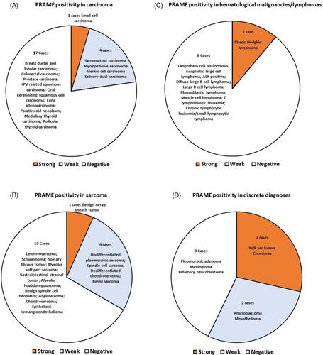
PRAME was detected in tumor cells in 86% of melanoma cases, which was significantly lower than SOX10 (100%) (p < .01), and similar to HMB45 (84%) and Melan-A (82%). S-100 had the lowest sensitivity of 71%. In comparison to other types of melanomas, spindle cell melanoma exhibited higher negativity for PRAME IHC (4/10 = 40%). PRAME was also expressed in some nonmelanocytic malignancies including carcinoma (5/22 = 23%), sarcoma (5/15 = 33%), and hematologic malignancies (1/9 = 11%). Overall, PRAME showed a sensitivity of 86%, specificity of 82%, positive predictive value of 70%, and negative predictive value of 92% for metastatic melanoma.
Conclusions
PRAME is a useful marker for the diagnosis of melanoma in cytology material, but it is less sensitive than SOX10. PRAME is also expressed in other nonmelanocytic tumors which limits its specificity.
期刊介绍:
Diagnostic Cytopathology is intended to provide a forum for the exchange of information in the field of cytopathology, with special emphasis on the practical, clinical aspects of the discipline. The editors invite original scientific articles, as well as special review articles, feature articles, and letters to the editor, from laboratory professionals engaged in the practice of cytopathology. Manuscripts are accepted for publication on the basis of scientific merit, practical significance, and suitability for publication in a journal dedicated to this discipline. Original articles can be considered only with the understanding that they have never been published before and that they have not been submitted for simultaneous review to another publication.


 分享
分享
 求助内容:
求助内容: 应助结果提醒方式:
 扫码关注我们
