Elizabeth Lea Schmidt, Zihao Ou, Erving Ximendes, Han Cui, Carl H. C. Keck, Daniel Jaque, Guosong Hong
{"title":"Near-infrared II fluorescence imaging","authors":"Elizabeth Lea Schmidt, Zihao Ou, Erving Ximendes, Han Cui, Carl H. C. Keck, Daniel Jaque, Guosong Hong","doi":"10.1038/s43586-024-00301-x","DOIUrl":null,"url":null,"abstract":"Fluorescence imaging in the second near-infrared (NIR-II) window enables deep-tissue imaging with high resolution and improved contrast by taking advantage of the reduced light scattering and tissue autofluorescence in this region of the spectrum. NIR-II fluorescence imaging uses photoluminescent contrast agents — including carbon nanotubes, quantum dots, rare earth-doped nanocrystals, gold nanoclusters, small molecules and their aggregates — and fluorescent proteins, which all exhibit fluorescence in the 1,000–3,000 nm range. After administration of these fluorophores in vivo, live animals can be imaged with specialized detectors and optical instruments, yielding images with contrast and resolution unparalleled by conventional visible and near-infrared fluorescence imaging. This powerful approach enables dynamic imaging of vascular structures and haemodynamics; molecular imaging and image-guided surgery of tumours; and visualization of deep-seated structures, such as the gastrointestinal system. NIR-II fluorescence imaging has revolutionized biomedical imaging over the past 15 years and is poised to make comparable advancements in cardiology, neurobiology and gastroenterology. This Primer describes the principles of NIR-II fluorescence imaging, reviews the most used fluorophores, outlines implementation approaches and discusses specific scientific and clinical applications. Furthermore, the limitations of NIR-II fluorescence imaging are addressed and future opportunities across various scientific domains are explored. Deep tissues can be imaged with high resolution and greater contrast by performing fluorescence imaging in the second near-infrared (NIR-II) window. This Primer summarizes how NIR-II fluorescence imaging can be used in animal models, exploring commonly used fluorophores and implementation approaches across a range of scientific and clinical applications.","PeriodicalId":74250,"journal":{"name":"Nature reviews. Methods primers","volume":" ","pages":"1-22"},"PeriodicalIF":56.0000,"publicationDate":"2024-04-04","publicationTypes":"Journal Article","fieldsOfStudy":null,"isOpenAccess":false,"openAccessPdf":"","citationCount":"0","resultStr":null,"platform":"Semanticscholar","paperid":null,"PeriodicalName":"Nature reviews. Methods primers","FirstCategoryId":"1085","ListUrlMain":"https://www.nature.com/articles/s43586-024-00301-x","RegionNum":0,"RegionCategory":null,"ArticlePicture":[],"TitleCN":null,"AbstractTextCN":null,"PMCID":null,"EPubDate":"","PubModel":"","JCR":"Q1","JCRName":"MULTIDISCIPLINARY SCIENCES","Score":null,"Total":0}
引用次数: 0
Abstract
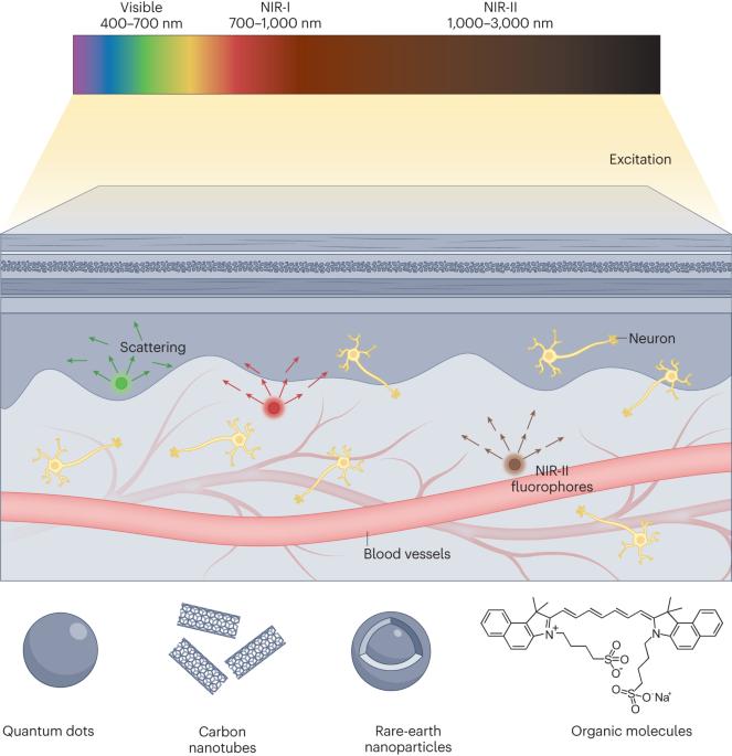
Fluorescence imaging in the second near-infrared (NIR-II) window enables deep-tissue imaging with high resolution and improved contrast by taking advantage of the reduced light scattering and tissue autofluorescence in this region of the spectrum. NIR-II fluorescence imaging uses photoluminescent contrast agents — including carbon nanotubes, quantum dots, rare earth-doped nanocrystals, gold nanoclusters, small molecules and their aggregates — and fluorescent proteins, which all exhibit fluorescence in the 1,000–3,000 nm range. After administration of these fluorophores in vivo, live animals can be imaged with specialized detectors and optical instruments, yielding images with contrast and resolution unparalleled by conventional visible and near-infrared fluorescence imaging. This powerful approach enables dynamic imaging of vascular structures and haemodynamics; molecular imaging and image-guided surgery of tumours; and visualization of deep-seated structures, such as the gastrointestinal system. NIR-II fluorescence imaging has revolutionized biomedical imaging over the past 15 years and is poised to make comparable advancements in cardiology, neurobiology and gastroenterology. This Primer describes the principles of NIR-II fluorescence imaging, reviews the most used fluorophores, outlines implementation approaches and discusses specific scientific and clinical applications. Furthermore, the limitations of NIR-II fluorescence imaging are addressed and future opportunities across various scientific domains are explored. Deep tissues can be imaged with high resolution and greater contrast by performing fluorescence imaging in the second near-infrared (NIR-II) window. This Primer summarizes how NIR-II fluorescence imaging can be used in animal models, exploring commonly used fluorophores and implementation approaches across a range of scientific and clinical applications.


 分享
分享

 求助内容:
求助内容: 应助结果提醒方式:
 扫码关注我们
