Yulia M. Alexandrovskaya, Ekaterina M. Kasianenko, Alexander A. Sovetsky, Alexander L. Matveyev, Dmitry A. Atyakshin, Olga I. Patsap, Mikhail A. Ignatiuk, Artem V. Volodkin, Vladimir Y. Zaitsev
{"title":"Optical coherence elastography with osmotically induced strains: Preliminary demonstration for express detection of cartilage degradation","authors":"Yulia M. Alexandrovskaya, Ekaterina M. Kasianenko, Alexander A. Sovetsky, Alexander L. Matveyev, Dmitry A. Atyakshin, Olga I. Patsap, Mikhail A. Ignatiuk, Artem V. Volodkin, Vladimir Y. Zaitsev","doi":"10.1002/jbio.202400016","DOIUrl":null,"url":null,"abstract":"<p>Optical coherence elastography (OCE) demonstrated impressive abilities for diagnosing tissue types/states using differences in their biomechanics. Usually, OCE visualizes tissue deformation induced by some additional stimulus (e.g., contact compression or auxiliary elastic-wave excitation). We propose a new variant of OCE with osmotically induced straining (OIS-OCE) and demonstrate its application to assess various stages of proteoglycan content degradation in cartilage. The information-bearing signatures in OIS-OCE are the magnitude and rate of strains caused by the application of osmotically active solutions onto the sample surface. OCE examination of the induced strains does not require special tissue preparation, the osmotic stimulation is highly reproducible, and strains are observed in noncontact mode. Several minutes suffice to obtain a conclusion. These features are promising for intraoperative method usage when express assessment of tissue state is required during surgical operations. The “waterfall” images demonstrate the development of cumulative osmotic strains in control and degraded cartilage samples.</p>","PeriodicalId":184,"journal":{"name":"Journal of Biophotonics","volume":"17 7","pages":""},"PeriodicalIF":2.0000,"publicationDate":"2024-05-04","publicationTypes":"Journal Article","fieldsOfStudy":null,"isOpenAccess":false,"openAccessPdf":"","citationCount":"0","resultStr":null,"platform":"Semanticscholar","paperid":null,"PeriodicalName":"Journal of Biophotonics","FirstCategoryId":"101","ListUrlMain":"https://onlinelibrary.wiley.com/doi/10.1002/jbio.202400016","RegionNum":3,"RegionCategory":"物理与天体物理","ArticlePicture":[],"TitleCN":null,"AbstractTextCN":null,"PMCID":null,"EPubDate":"","PubModel":"","JCR":"Q3","JCRName":"BIOCHEMICAL RESEARCH METHODS","Score":null,"Total":0}
引用次数: 0
Abstract
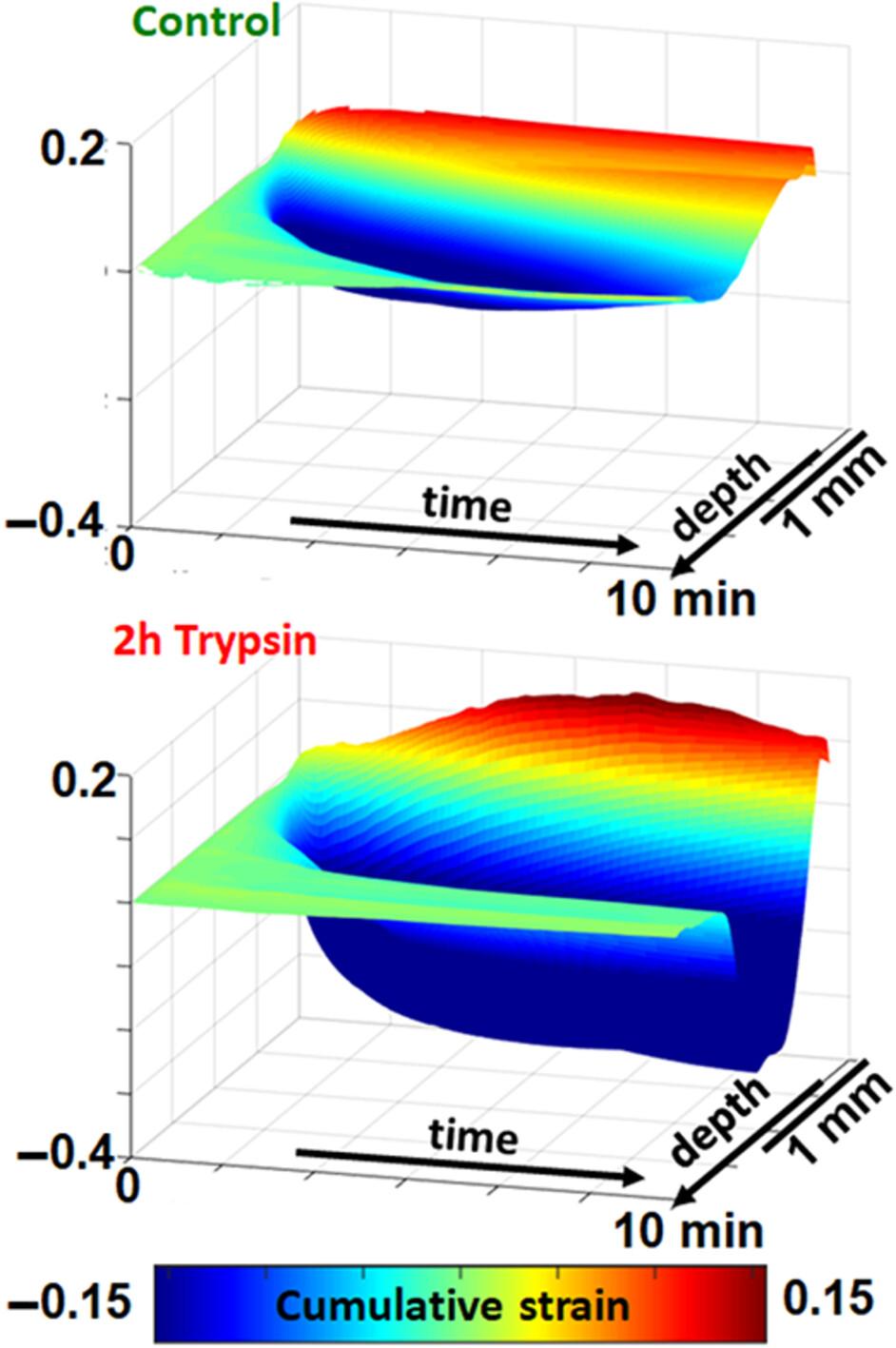
Optical coherence elastography (OCE) demonstrated impressive abilities for diagnosing tissue types/states using differences in their biomechanics. Usually, OCE visualizes tissue deformation induced by some additional stimulus (e.g., contact compression or auxiliary elastic-wave excitation). We propose a new variant of OCE with osmotically induced straining (OIS-OCE) and demonstrate its application to assess various stages of proteoglycan content degradation in cartilage. The information-bearing signatures in OIS-OCE are the magnitude and rate of strains caused by the application of osmotically active solutions onto the sample surface. OCE examination of the induced strains does not require special tissue preparation, the osmotic stimulation is highly reproducible, and strains are observed in noncontact mode. Several minutes suffice to obtain a conclusion. These features are promising for intraoperative method usage when express assessment of tissue state is required during surgical operations. The “waterfall” images demonstrate the development of cumulative osmotic strains in control and degraded cartilage samples.
期刊介绍:
The first international journal dedicated to publishing reviews and original articles from this exciting field, the Journal of Biophotonics covers the broad range of research on interactions between light and biological material. The journal offers a platform where the physicist communicates with the biologist and where the clinical practitioner learns about the latest tools for the diagnosis of diseases. As such, the journal is highly interdisciplinary, publishing cutting edge research in the fields of life sciences, medicine, physics, chemistry, and engineering. The coverage extends from fundamental research to specific developments, while also including the latest applications.


 分享
分享
 求助内容:
求助内容: 应助结果提醒方式:
 扫码关注我们
