Adrienn Tóth, Jordan H Chamberlin, Salvador Mendez, Akos Varga-Szemes, Andrew D Hardie
{"title":"Iodine quantification of renal lesions: Preliminary results using spectral-based material extraction on photon-counting CT.","authors":"Adrienn Tóth, Jordan H Chamberlin, Salvador Mendez, Akos Varga-Szemes, Andrew D Hardie","doi":"10.25259/JCIS_1_2024","DOIUrl":null,"url":null,"abstract":"<p><strong>Objectives: </strong>To assess the range of quantitative iodine values in renal cysts (RC) (with a few renal neoplasms [RNs] as a comparison) to develop an expected range of values for RC that can be used in future studies for their differentiation.</p><p><strong>Material and methods: </strong>Consecutive patients (<i>n</i> = 140) with renal lesions who had undergone abdominal examination on a clinical photon-counting computed tomography (PCCT) were retrospectively included. Automated iodine quantification maps were reconstructed, and region of interest (ROI) measurements of iodine concentration (IC) (mg/cm<sup>3</sup>) were performed on whole renal lesions. In addition, for heterogeneous lesions, a secondary ROI was placed on the area most suspicious for malignancy. The discriminatory values of minimum, maximum, mean, and standard deviation for IC were compared using simple logistic regression and receiver operating characteristic curves (area under the curve [AUC]).</p><p><strong>Results: </strong>A total of 259 renal lesions (243 RC and 16 RN) were analyzed. There were significant differences between RC and RN for all IC measures with the best-performing metrics being mean and maximum IC of the entire lesion ROI (AUC 0.912 and 0.917, respectively) but also mean and minimum IC of the most suspicious area in heterogeneous lesions (AUC 0.983 and 0.992, respectively). Most RC fell within a range of low measured iodine values although a few had higher values.</p><p><strong>Conclusion: </strong>Automated iodine quantification maps reconstructed from clinical PCCT have a high diagnostic ability to differentiate RCs and neoplasms. The data from this pilot study can be used to help establish quantitative values for clinical differentiation of renal lesions.</p>","PeriodicalId":15512,"journal":{"name":"Journal of Clinical Imaging Science","volume":"14 ","pages":"7"},"PeriodicalIF":1.3000,"publicationDate":"2024-03-08","publicationTypes":"Journal Article","fieldsOfStudy":null,"isOpenAccess":false,"openAccessPdf":"https://www.ncbi.nlm.nih.gov/pmc/articles/PMC11021115/pdf/","citationCount":"0","resultStr":null,"platform":"Semanticscholar","paperid":null,"PeriodicalName":"Journal of Clinical Imaging Science","FirstCategoryId":"1085","ListUrlMain":"https://doi.org/10.25259/JCIS_1_2024","RegionNum":0,"RegionCategory":null,"ArticlePicture":[],"TitleCN":null,"AbstractTextCN":null,"PMCID":null,"EPubDate":"2024/1/1 0:00:00","PubModel":"eCollection","JCR":"Q3","JCRName":"RADIOLOGY, NUCLEAR MEDICINE & MEDICAL IMAGING","Score":null,"Total":0}
引用次数: 0
Abstract
Objectives: To assess the range of quantitative iodine values in renal cysts (RC) (with a few renal neoplasms [RNs] as a comparison) to develop an expected range of values for RC that can be used in future studies for their differentiation.
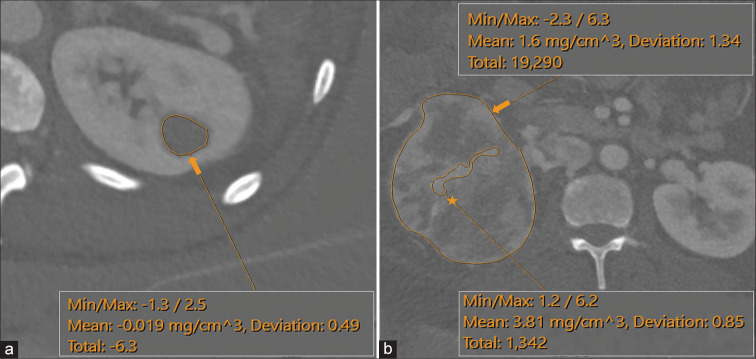
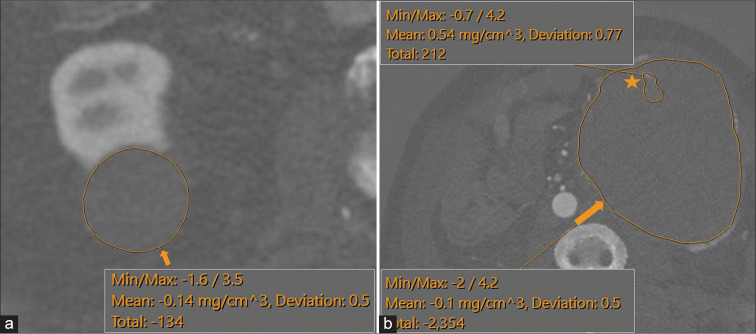
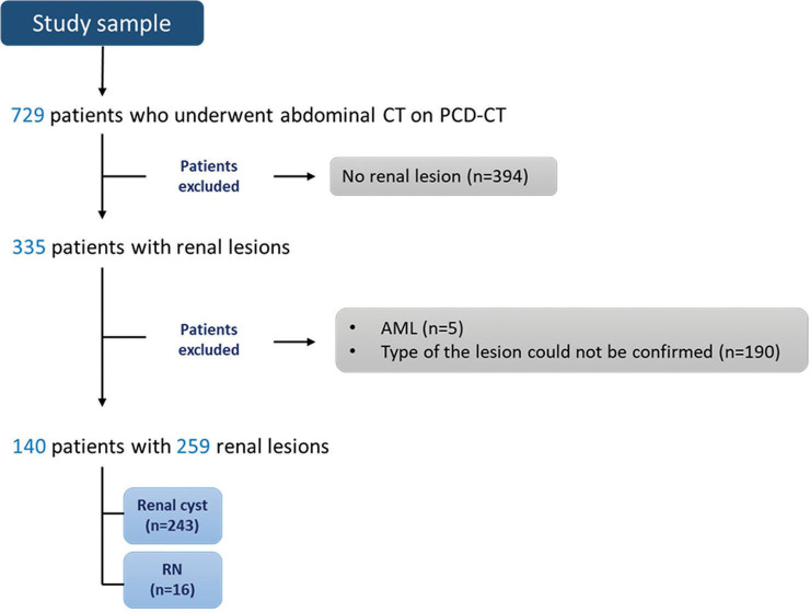
Material and methods: Consecutive patients (n = 140) with renal lesions who had undergone abdominal examination on a clinical photon-counting computed tomography (PCCT) were retrospectively included. Automated iodine quantification maps were reconstructed, and region of interest (ROI) measurements of iodine concentration (IC) (mg/cm3) were performed on whole renal lesions. In addition, for heterogeneous lesions, a secondary ROI was placed on the area most suspicious for malignancy. The discriminatory values of minimum, maximum, mean, and standard deviation for IC were compared using simple logistic regression and receiver operating characteristic curves (area under the curve [AUC]).
Results: A total of 259 renal lesions (243 RC and 16 RN) were analyzed. There were significant differences between RC and RN for all IC measures with the best-performing metrics being mean and maximum IC of the entire lesion ROI (AUC 0.912 and 0.917, respectively) but also mean and minimum IC of the most suspicious area in heterogeneous lesions (AUC 0.983 and 0.992, respectively). Most RC fell within a range of low measured iodine values although a few had higher values.
Conclusion: Automated iodine quantification maps reconstructed from clinical PCCT have a high diagnostic ability to differentiate RCs and neoplasms. The data from this pilot study can be used to help establish quantitative values for clinical differentiation of renal lesions.
期刊介绍:
The Journal of Clinical Imaging Science (JCIS) is an open access peer-reviewed journal committed to publishing high-quality articles in the field of Imaging Science. The journal aims to present Imaging Science and relevant clinical information in an understandable and useful format. The journal is owned and published by the Scientific Scholar. Audience Our audience includes Radiologists, Researchers, Clinicians, medical professionals and students. Review process JCIS has a highly rigorous peer-review process that makes sure that manuscripts are scientifically accurate, relevant, novel and important. Authors disclose all conflicts, affiliations and financial associations such that the published content is not biased.


 分享
分享
 求助内容:
求助内容: 应助结果提醒方式:
 扫码关注我们
