Kartik Viswanathan, Elan Hahn, Snjezana Dogan, Ilan Weinreb, Brendan C Dickson, Christina MacMillan, Nora Katabi, Kelly Magliocca, Ronald Ghossein, Bin Xu
{"title":"The histological spectrum and immunoprofile of head and neck NUT carcinoma: A multicentre series of 30 cases","authors":"Kartik Viswanathan, Elan Hahn, Snjezana Dogan, Ilan Weinreb, Brendan C Dickson, Christina MacMillan, Nora Katabi, Kelly Magliocca, Ronald Ghossein, Bin Xu","doi":"10.1111/his.15204","DOIUrl":null,"url":null,"abstract":"<div>\n \n <section>\n \n <h3> Background and aim</h3>\n \n <p>Head and neck nuclear protein of testis carcinoma (HN-NUT) is a rare form of carcinoma diagnosed by NUT immunohistochemistry positivity and/or <i>NUTM1</i> translocation. Although the prototype of HN-NUT is a primitive undifferentiated round cell tumour (URC) with immunopositivity for squamous markers, it is our observation that it may assume variant histology or immunoprofile.</p>\n </section>\n \n <section>\n \n <h3> Methods</h3>\n \n <p>We conducted a detailed clinicopathological review of a large retrospective cohort of 30 HN-NUT, aiming to expand its histological and immunohistochemical spectrum.</p>\n </section>\n \n <section>\n \n <h3> Results</h3>\n \n <p>The median age of patients with HN-NUT was 39 years (range = 17–86). It affected the sinonasal tract (43%), major salivary glands (20%), thyroid (13%), oral cavity (7%), larynx (7%), neck (7%) and nasopharynx (3%). Although most cases of HN-NUT (63%) contained a component of primitive URC tumour, 53% showed other histological features and 37% lacked a URC component altogether. Variant histological features included basaloid (33%), differentiated squamous/squamoid (37%), clear cell changes (13%), glandular differentiation (7%) and papillary architecture (10%), which could co-exist. While most HN-NUT were positive for keratins, p63 and p40, occasional cases (5–9%) were entirely negative. Immunopositivity for neuroendocrine markers and thyroid transcription factor-1 was observed in 33 and 36% of cases, respectively. The outcome of HN-NUT was dismal, with a 3-year disease specific survival of 38%.</p>\n </section>\n \n <section>\n \n <h3> Conclusions</h3>\n \n <p>HN-NUT can affect individuals across a wide age range and arise from various head and neck sites. It exhibits a diverse spectrum of histological features and may be positive for neuroendocrine markers, potentially leading to underdiagnosis. A low threshold to perform NUT-specific tests is necessary to accurately diagnose HN-NUT.</p>\n </section>\n </div>","PeriodicalId":13219,"journal":{"name":"Histopathology","volume":"85 2","pages":"317-326"},"PeriodicalIF":4.1000,"publicationDate":"2024-05-06","publicationTypes":"Journal Article","fieldsOfStudy":null,"isOpenAccess":false,"openAccessPdf":"","citationCount":"0","resultStr":null,"platform":"Semanticscholar","paperid":null,"PeriodicalName":"Histopathology","FirstCategoryId":"3","ListUrlMain":"https://onlinelibrary.wiley.com/doi/10.1111/his.15204","RegionNum":2,"RegionCategory":"医学","ArticlePicture":[],"TitleCN":null,"AbstractTextCN":null,"PMCID":null,"EPubDate":"","PubModel":"","JCR":"Q2","JCRName":"CELL BIOLOGY","Score":null,"Total":0}
引用次数: 0
Abstract
Background and aim
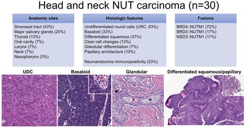
Head and neck nuclear protein of testis carcinoma (HN-NUT) is a rare form of carcinoma diagnosed by NUT immunohistochemistry positivity and/or NUTM1 translocation. Although the prototype of HN-NUT is a primitive undifferentiated round cell tumour (URC) with immunopositivity for squamous markers, it is our observation that it may assume variant histology or immunoprofile.
Methods
We conducted a detailed clinicopathological review of a large retrospective cohort of 30 HN-NUT, aiming to expand its histological and immunohistochemical spectrum.
Results
The median age of patients with HN-NUT was 39 years (range = 17–86). It affected the sinonasal tract (43%), major salivary glands (20%), thyroid (13%), oral cavity (7%), larynx (7%), neck (7%) and nasopharynx (3%). Although most cases of HN-NUT (63%) contained a component of primitive URC tumour, 53% showed other histological features and 37% lacked a URC component altogether. Variant histological features included basaloid (33%), differentiated squamous/squamoid (37%), clear cell changes (13%), glandular differentiation (7%) and papillary architecture (10%), which could co-exist. While most HN-NUT were positive for keratins, p63 and p40, occasional cases (5–9%) were entirely negative. Immunopositivity for neuroendocrine markers and thyroid transcription factor-1 was observed in 33 and 36% of cases, respectively. The outcome of HN-NUT was dismal, with a 3-year disease specific survival of 38%.
Conclusions
HN-NUT can affect individuals across a wide age range and arise from various head and neck sites. It exhibits a diverse spectrum of histological features and may be positive for neuroendocrine markers, potentially leading to underdiagnosis. A low threshold to perform NUT-specific tests is necessary to accurately diagnose HN-NUT.
期刊介绍:
Histopathology is an international journal intended to be of practical value to surgical and diagnostic histopathologists, and to investigators of human disease who employ histopathological methods. Our primary purpose is to publish advances in pathology, in particular those applicable to clinical practice and contributing to the better understanding of human disease.


 分享
分享
 求助内容:
求助内容: 应助结果提醒方式:
 扫码关注我们
