Hisaki Aiba, Alberto Righi, Paolo Spinnato, Alessandra Longhi, Giorgio Frega, Ahmed Atherley O'Meally, Ayano Aso, Konstantina Solou, Barbara Dozza, Marco Gambarotti, Toni Ibrahim, Davide Maria Donati, Costantino Errani
{"title":"Histological and imaging features of myoepithelial carcinoma of the bone and soft tissue.","authors":"Hisaki Aiba, Alberto Righi, Paolo Spinnato, Alessandra Longhi, Giorgio Frega, Ahmed Atherley O'Meally, Ayano Aso, Konstantina Solou, Barbara Dozza, Marco Gambarotti, Toni Ibrahim, Davide Maria Donati, Costantino Errani","doi":"10.1007/s00256-024-04693-5","DOIUrl":null,"url":null,"abstract":"<p><strong>Objective: </strong>To depict histological and imaging features of myoepithelial carcinoma of the bone and soft tissue.</p><p><strong>Materials and methods: </strong>We retrospectively examined histological features in 22 patients with myoepithelial carcinoma of the bone (4 patients) and soft tissue (18 patients) at a single institution. Imaging analysis of 15 patients (bone, 3 patients; soft tissue, 12 patients;) with preoperative images involved classifying lytic bone lesions via the modified Lodwick-Madewell classification; the growth patterns of soft tissue lesions were classified as well-defined, focally invasive, or diffusely invasive.</p><p><strong>Results: </strong>Local recurrence occurred in eight out of 22 patients (36.3%). Four of 22 patients (18.2%) had metastasis at presentation, whereas 11 of 22 patients (50.0%) had distant metastasis during follow-up. Severe cytological pleomorphism was observed in 14 of 22 patients (63.6%), and 10 of 22 tumors (45.5%) showed ≥ 10 mitoses/10 high-power fields. Vascular invasion was observed in 10 of 22 patients (45.5%). Extracapsular/extraskeletal infiltration into the surrounding tissues was assessed in 20 patients, with 14 of them (70%) showing infiltration beyond the tumor border. Regarding imaging of bone lesions, two patients had Ludwick type IIIB, whereas one patient had type II. The growth pattern of soft tissue lesions was well-defined in two patients (16.7%), focally invasive in seven patients (58.3%), and diffusely invasive in three (25.0%) out of 12 patients.</p><p><strong>Conclusion: </strong>Myoepithelial carcinoma of the bone and soft tissue presents high risk of local recurrence and distant metastasis. Histological and imaging features might be important to understand the aggressive behavior of the tumor.</p>","PeriodicalId":21783,"journal":{"name":"Skeletal Radiology","volume":" ","pages":"2617-2625"},"PeriodicalIF":2.2000,"publicationDate":"2024-12-01","publicationTypes":"Journal Article","fieldsOfStudy":null,"isOpenAccess":false,"openAccessPdf":"","citationCount":"0","resultStr":null,"platform":"Semanticscholar","paperid":null,"PeriodicalName":"Skeletal Radiology","FirstCategoryId":"3","ListUrlMain":"https://doi.org/10.1007/s00256-024-04693-5","RegionNum":3,"RegionCategory":"医学","ArticlePicture":[],"TitleCN":null,"AbstractTextCN":null,"PMCID":null,"EPubDate":"2024/4/29 0:00:00","PubModel":"Epub","JCR":"Q2","JCRName":"ORTHOPEDICS","Score":null,"Total":0}
引用次数: 0
Abstract
Objective: To depict histological and imaging features of myoepithelial carcinoma of the bone and soft tissue.
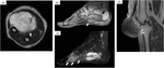
Materials and methods: We retrospectively examined histological features in 22 patients with myoepithelial carcinoma of the bone (4 patients) and soft tissue (18 patients) at a single institution. Imaging analysis of 15 patients (bone, 3 patients; soft tissue, 12 patients;) with preoperative images involved classifying lytic bone lesions via the modified Lodwick-Madewell classification; the growth patterns of soft tissue lesions were classified as well-defined, focally invasive, or diffusely invasive.
Results: Local recurrence occurred in eight out of 22 patients (36.3%). Four of 22 patients (18.2%) had metastasis at presentation, whereas 11 of 22 patients (50.0%) had distant metastasis during follow-up. Severe cytological pleomorphism was observed in 14 of 22 patients (63.6%), and 10 of 22 tumors (45.5%) showed ≥ 10 mitoses/10 high-power fields. Vascular invasion was observed in 10 of 22 patients (45.5%). Extracapsular/extraskeletal infiltration into the surrounding tissues was assessed in 20 patients, with 14 of them (70%) showing infiltration beyond the tumor border. Regarding imaging of bone lesions, two patients had Ludwick type IIIB, whereas one patient had type II. The growth pattern of soft tissue lesions was well-defined in two patients (16.7%), focally invasive in seven patients (58.3%), and diffusely invasive in three (25.0%) out of 12 patients.
Conclusion: Myoepithelial carcinoma of the bone and soft tissue presents high risk of local recurrence and distant metastasis. Histological and imaging features might be important to understand the aggressive behavior of the tumor.
期刊介绍:
Skeletal Radiology provides a forum for the dissemination of current knowledge and information dealing with disorders of the musculoskeletal system including the spine. While emphasizing the radiological aspects of the many varied skeletal abnormalities, the journal also adopts an interdisciplinary approach, reflecting the membership of the International Skeletal Society. Thus, the anatomical, pathological, physiological, clinical, metabolic and epidemiological aspects of the many entities affecting the skeleton receive appropriate consideration.
This is the Journal of the International Skeletal Society and the Official Journal of the Society of Skeletal Radiology and the Australasian Musculoskelelal Imaging Group.


 分享
分享
 求助内容:
求助内容: 应助结果提醒方式:
 扫码关注我们
