{"title":"Utility of intraoral scanner imaging for dental plaque detection.","authors":"Chihiro Yoshiga, Kazuya Doi, Hiroshi Oue, Reiko Kobatake, Maiko Kawagoe, Hanako Umehara, Kazuhiro Tsuga","doi":"10.5624/isd.20230180","DOIUrl":null,"url":null,"abstract":"<p><strong>Purpose: </strong>Oral hygiene, maintained through plaque control, helps prevent periodontal disease and dental caries. This study was conducted to examine the accuracy of plaque detection with an intraoral scanner (IOS) compared to images captured with an optical camera.</p><p><strong>Materials and methods: </strong>To examine the effect of color tone, artificial tooth resin samples were stained red, blue, and green, after which images were acquired with a digital single-lens reflex (DSLR) camera and an IOS device. Stained surface ratios were then determined and compared. Additionally, the deviation rate of the IOS relative to the DSLR camera was computed for each color. In the clinical study, following plaque staining with red disclosing solution, the staining was captured by the DSLR and IOS devices, and the stained area on each image was measured.</p><p><strong>Results: </strong>The stained surface ratios did not differ significantly between DSLR and IOS images for any color group. Additionally, the deviation rate did not vary significantly across colors. In the clinical test, the stained plaque appeared slightly lighter in color, and the delineation of the stained areas less distinct, on the IOS compared to the DSLR images. However, the stained surface ratio was significantly higher in the IOS than in the DSLR group.</p><p><strong>Conclusion: </strong>When employing IOS with dental plaque staining, the impact of color was minimal, suggesting that the traditional red stain remains suitable for plaque detection. IOS images appeared relatively blurred and enlarged relative to the true state of the teeth, due to inferior sharpness compared to camera images.</p>","PeriodicalId":51714,"journal":{"name":"Imaging Science in Dentistry","volume":"54 1","pages":"43-48"},"PeriodicalIF":2.1000,"publicationDate":"2024-03-01","publicationTypes":"Journal Article","fieldsOfStudy":null,"isOpenAccess":false,"openAccessPdf":"https://www.ncbi.nlm.nih.gov/pmc/articles/PMC10985524/pdf/","citationCount":"0","resultStr":null,"platform":"Semanticscholar","paperid":null,"PeriodicalName":"Imaging Science in Dentistry","FirstCategoryId":"1085","ListUrlMain":"https://doi.org/10.5624/isd.20230180","RegionNum":0,"RegionCategory":null,"ArticlePicture":[],"TitleCN":null,"AbstractTextCN":null,"PMCID":null,"EPubDate":"2024/1/4 0:00:00","PubModel":"Epub","JCR":"Q3","JCRName":"DENTISTRY, ORAL SURGERY & MEDICINE","Score":null,"Total":0}
引用次数: 0
Abstract
Purpose: Oral hygiene, maintained through plaque control, helps prevent periodontal disease and dental caries. This study was conducted to examine the accuracy of plaque detection with an intraoral scanner (IOS) compared to images captured with an optical camera.
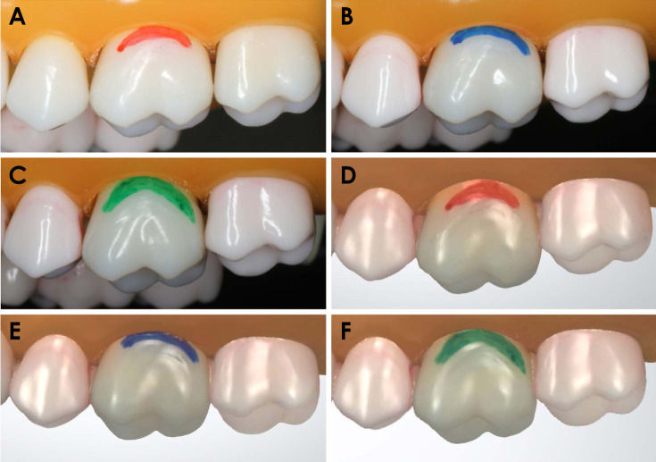
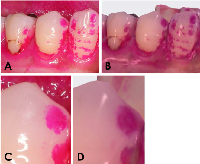
Materials and methods: To examine the effect of color tone, artificial tooth resin samples were stained red, blue, and green, after which images were acquired with a digital single-lens reflex (DSLR) camera and an IOS device. Stained surface ratios were then determined and compared. Additionally, the deviation rate of the IOS relative to the DSLR camera was computed for each color. In the clinical study, following plaque staining with red disclosing solution, the staining was captured by the DSLR and IOS devices, and the stained area on each image was measured.
Results: The stained surface ratios did not differ significantly between DSLR and IOS images for any color group. Additionally, the deviation rate did not vary significantly across colors. In the clinical test, the stained plaque appeared slightly lighter in color, and the delineation of the stained areas less distinct, on the IOS compared to the DSLR images. However, the stained surface ratio was significantly higher in the IOS than in the DSLR group.
Conclusion: When employing IOS with dental plaque staining, the impact of color was minimal, suggesting that the traditional red stain remains suitable for plaque detection. IOS images appeared relatively blurred and enlarged relative to the true state of the teeth, due to inferior sharpness compared to camera images.


 分享
分享
 求助内容:
求助内容: 应助结果提醒方式:
 扫码关注我们
