Dogan S Polat, Son Nguyen, Paniz Karbasi, Keith Hulsey, Murat Can Cobanoglu, Liqiang Wang, Albert Montillo, Basak E Dogan
{"title":"Machine Learning Prediction of Lymph Node Metastasis in Breast Cancer: Performance of a Multi-institutional MRI-based 4D Convolutional Neural Network.","authors":"Dogan S Polat, Son Nguyen, Paniz Karbasi, Keith Hulsey, Murat Can Cobanoglu, Liqiang Wang, Albert Montillo, Basak E Dogan","doi":"10.1148/rycan.230107","DOIUrl":null,"url":null,"abstract":"<p><p>Purpose To develop a custom deep convolutional neural network (CNN) for noninvasive prediction of breast cancer nodal metastasis. Materials and Methods This retrospective study included patients with newly diagnosed primary invasive breast cancer with known pathologic (pN) and clinical nodal (cN) status who underwent dynamic contrast-enhanced (DCE) breast MRI at the authors' institution between July 2013 and July 2016. Clinicopathologic data (age, estrogen receptor and human epidermal growth factor 2 status, Ki-67 index, and tumor grade) and cN and pN status were collected. A four-dimensional (4D) CNN model integrating temporal information from dynamic image sets was developed. The convolutional layers learned prognostic image features, which were combined with clinicopathologic measures to predict cN0 versus cN+ and pN0 versus pN+ disease. Performance was assessed with the area under the receiver operating characteristic curve (AUC), with fivefold nested cross-validation. Results Data from 350 female patients (mean age, 51.7 years ± 11.9 [SD]) were analyzed. AUC, sensitivity, and specificity values of the 4D hybrid model were 0.87 (95% CI: 0.83, 0.91), 89% (95% CI: 79%, 93%), and 76% (95% CI: 68%, 88%) for differentiating pN0 versus pN+ and 0.79 (95% CI: 0.76, 0.82), 80% (95% CI: 77%, 84%), and 62% (95% CI: 58%, 67%), respectively, for differentiating cN0 versus cN+. Conclusion The proposed deep learning model using tumor DCE MR images demonstrated high sensitivity in identifying breast cancer lymph node metastasis and shows promise for potential use as a clinical decision support tool. <b>Keywords:</b> MR Imaging, Breast, Breast Cancer, Breast MRI, Machine Learning, Metastasis, Prognostic Prediction <i>Supplemental material is available for this article.</i> Published under a CC BY 4.0 license.</p>","PeriodicalId":20786,"journal":{"name":"Radiology. Imaging cancer","volume":"6 3","pages":"e230107"},"PeriodicalIF":5.6000,"publicationDate":"2024-05-01","publicationTypes":"Journal Article","fieldsOfStudy":null,"isOpenAccess":false,"openAccessPdf":"https://www.ncbi.nlm.nih.gov/pmc/articles/PMC11148663/pdf/","citationCount":"0","resultStr":null,"platform":"Semanticscholar","paperid":null,"PeriodicalName":"Radiology. Imaging cancer","FirstCategoryId":"1085","ListUrlMain":"https://doi.org/10.1148/rycan.230107","RegionNum":0,"RegionCategory":null,"ArticlePicture":[],"TitleCN":null,"AbstractTextCN":null,"PMCID":null,"EPubDate":"","PubModel":"","JCR":"Q1","JCRName":"ONCOLOGY","Score":null,"Total":0}
引用次数: 0
Abstract
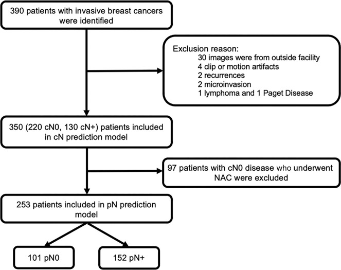
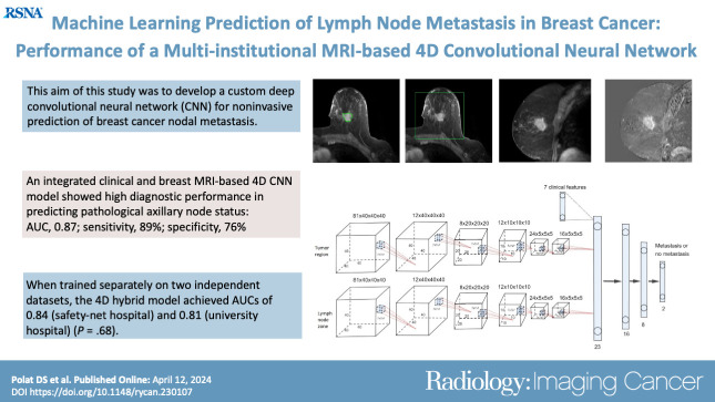
Purpose To develop a custom deep convolutional neural network (CNN) for noninvasive prediction of breast cancer nodal metastasis. Materials and Methods This retrospective study included patients with newly diagnosed primary invasive breast cancer with known pathologic (pN) and clinical nodal (cN) status who underwent dynamic contrast-enhanced (DCE) breast MRI at the authors' institution between July 2013 and July 2016. Clinicopathologic data (age, estrogen receptor and human epidermal growth factor 2 status, Ki-67 index, and tumor grade) and cN and pN status were collected. A four-dimensional (4D) CNN model integrating temporal information from dynamic image sets was developed. The convolutional layers learned prognostic image features, which were combined with clinicopathologic measures to predict cN0 versus cN+ and pN0 versus pN+ disease. Performance was assessed with the area under the receiver operating characteristic curve (AUC), with fivefold nested cross-validation. Results Data from 350 female patients (mean age, 51.7 years ± 11.9 [SD]) were analyzed. AUC, sensitivity, and specificity values of the 4D hybrid model were 0.87 (95% CI: 0.83, 0.91), 89% (95% CI: 79%, 93%), and 76% (95% CI: 68%, 88%) for differentiating pN0 versus pN+ and 0.79 (95% CI: 0.76, 0.82), 80% (95% CI: 77%, 84%), and 62% (95% CI: 58%, 67%), respectively, for differentiating cN0 versus cN+. Conclusion The proposed deep learning model using tumor DCE MR images demonstrated high sensitivity in identifying breast cancer lymph node metastasis and shows promise for potential use as a clinical decision support tool. Keywords: MR Imaging, Breast, Breast Cancer, Breast MRI, Machine Learning, Metastasis, Prognostic Prediction Supplemental material is available for this article. Published under a CC BY 4.0 license.


 分享
分享

 求助内容:
求助内容: 应助结果提醒方式:
 扫码关注我们
