Y. Zenmei Ohkubo , Peter W. Radulovic , Albert N. Kahira , Jesper J. Madsen
{"title":"Membrane binding and lipid-protein interaction of the C2 domain from coagulation factor V","authors":"Y. Zenmei Ohkubo , Peter W. Radulovic , Albert N. Kahira , Jesper J. Madsen","doi":"10.1016/j.crstbi.2024.100149","DOIUrl":null,"url":null,"abstract":"<div><p>Anchoring of coagulation factors to anionic regions of the membrane involves the C2 domain as a key player. The rate of enzymatic reactions of the coagulation factors is increased by several orders of magnitude upon membrane binding. However, the precise mechanisms behind the rate acceleration remain unclear, primarily because of a lack of understanding of the conformational dynamics of the C2-containing factors and corresponding complexes. We elucidate the membrane-bound form of the C2 domain from human coagulation factor V (FV–C2) by characterizing its membrane binding the specific lipid-protein interactions. Employing all-atom molecular dynamics simulations and leveraging the highly mobile membrane-mimetic (HMMM) model, we observed spontaneous binding of FV-C2 to a phosphatidylserine (PS)-containing membrane within 2–25 ns across twelve independent simulations. FV-C2 interacted with the membrane through three loops (spikes 1–3), achieving a converged, stable orientation. Multiple HMMM trajectories of the spontaneous membrane binding provided extensive sampling and ample data to examine the membrane-induced effects on the conformational dynamics of C2 as well as specific lipid-protein interactions. Despite existing crystal structures representing presumed “open” and “closed” states of FV-C2, our results revealed a continuous distribution of structures between these states, with the most populated structures differing from both “open” and “closed” states observed in crystal environments. Lastly, we characterized a putative PS-specific binding site formed by K23, Q48, and S78 located in the groove enclosed by spikes 1–3 (PS-specificity pocket), suggesting a different orientation of a bound headgroup moiety compared to previous proposals based upon analysis of static crystal structures.</p></div>","PeriodicalId":10870,"journal":{"name":"Current Research in Structural Biology","volume":"7 ","pages":"Article 100149"},"PeriodicalIF":2.7000,"publicationDate":"2024-01-01","publicationTypes":"Journal Article","fieldsOfStudy":null,"isOpenAccess":false,"openAccessPdf":"https://www.sciencedirect.com/science/article/pii/S2665928X24000266/pdfft?md5=e8ab6e6a13338f75043488820ca37b17&pid=1-s2.0-S2665928X24000266-main.pdf","citationCount":"0","resultStr":null,"platform":"Semanticscholar","paperid":null,"PeriodicalName":"Current Research in Structural Biology","FirstCategoryId":"1085","ListUrlMain":"https://www.sciencedirect.com/science/article/pii/S2665928X24000266","RegionNum":0,"RegionCategory":null,"ArticlePicture":[],"TitleCN":null,"AbstractTextCN":null,"PMCID":null,"EPubDate":"2024/5/1 0:00:00","PubModel":"Epub","JCR":"Q3","JCRName":"BIOCHEMISTRY & MOLECULAR BIOLOGY","Score":null,"Total":0}
引用次数: 0
Abstract
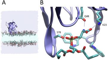
Anchoring of coagulation factors to anionic regions of the membrane involves the C2 domain as a key player. The rate of enzymatic reactions of the coagulation factors is increased by several orders of magnitude upon membrane binding. However, the precise mechanisms behind the rate acceleration remain unclear, primarily because of a lack of understanding of the conformational dynamics of the C2-containing factors and corresponding complexes. We elucidate the membrane-bound form of the C2 domain from human coagulation factor V (FV–C2) by characterizing its membrane binding the specific lipid-protein interactions. Employing all-atom molecular dynamics simulations and leveraging the highly mobile membrane-mimetic (HMMM) model, we observed spontaneous binding of FV-C2 to a phosphatidylserine (PS)-containing membrane within 2–25 ns across twelve independent simulations. FV-C2 interacted with the membrane through three loops (spikes 1–3), achieving a converged, stable orientation. Multiple HMMM trajectories of the spontaneous membrane binding provided extensive sampling and ample data to examine the membrane-induced effects on the conformational dynamics of C2 as well as specific lipid-protein interactions. Despite existing crystal structures representing presumed “open” and “closed” states of FV-C2, our results revealed a continuous distribution of structures between these states, with the most populated structures differing from both “open” and “closed” states observed in crystal environments. Lastly, we characterized a putative PS-specific binding site formed by K23, Q48, and S78 located in the groove enclosed by spikes 1–3 (PS-specificity pocket), suggesting a different orientation of a bound headgroup moiety compared to previous proposals based upon analysis of static crystal structures.


 分享
分享
 求助内容:
求助内容: 应助结果提醒方式:
 扫码关注我们
