{"title":"Ultrastructural Features of the Alimentary Canal in Hermaphroditic Appendicularians Oikopleura gracilis (Tunicata, Oikopleuridae)","authors":"A. V. Savelieva","doi":"10.1134/s1063074023080084","DOIUrl":null,"url":null,"abstract":"<h3 data-test=\"abstract-sub-heading\">Abstract</h3><p>Appendicularians play an important role in marine food webs, but the alimentary canal of hermaphroditic oikopleurids, which prevalent among urochordates, is little studied ultrastructurally. Serial microtomy revealed that the alimentary canal of <i>Oikopleura gracilis</i> consists of a wide pharynx and a U-shaped gut with typical for oikopleurids anatomy: oesophagus, a bilobed stomach, the vertical, short mid- and voluminous distal or rectal intestines, ending the anal papilla. The mid-intestine leaves under the right gastric lobe. TEM analysis diagnosed the following features in the alimentary canal of <i>Oikopleura gracilis</i>. First, the pharynx is lined with a single layer of highly flattened non-ciliated epithelium. Above the anterior part of the endostyle the pharyngeal epithelium forms a thickening – ciliated ring. It is formed by cells of two types: prismatic, ciliated microvillar cells arranged in several parallel rows, and two rows of narrow parietal cells covering the first cell type. Ciliated ring serves as a kind of first valve, prevents food reflux. Second, in the pharynx of one of the specimens three flagellate protozoans with ultrastructurally similarities of free-living kinetoplastids were found. Third, an additional row of gastric band cells was revealed in the upper part of the right gastric lobe compared to that of the gastric floor. Giant electron-lucent vesicles with homogenous content associated with rough endoplasmic reticulum found in the cytoplasm of the gastric band cell, indicating probable storage the protein digestive secrets in these cells. Large multivesicular bodies diagnosed in gastric band cells ultrastructurally corresponded to autophagosomes. Forth, a new cellular type was found, the osmiophilic enterocytes, scattered throughout the digestive epithelium, that shared ultrastructural signs with apoptotic cells. Fifth, TEM data revealed the peritrophic membrane structure and proposed its formation. The absence of its own microflora in the peritrophic membrane, i.e., the absence of an immunity barrier, may be connected with the short ontogeny of appendicularians. The ultrastructural features revealed in the alimentary canal of <i>O. gracilis</i> expand our knowledge of the functioning of the appendicularians and serve as the basis for further research.</p>","PeriodicalId":49584,"journal":{"name":"Russian Journal of Marine Biology","volume":"57 1","pages":""},"PeriodicalIF":0.4000,"publicationDate":"2024-01-29","publicationTypes":"Journal Article","fieldsOfStudy":null,"isOpenAccess":false,"openAccessPdf":"","citationCount":"0","resultStr":null,"platform":"Semanticscholar","paperid":null,"PeriodicalName":"Russian Journal of Marine Biology","FirstCategoryId":"99","ListUrlMain":"https://doi.org/10.1134/s1063074023080084","RegionNum":4,"RegionCategory":"生物学","ArticlePicture":[],"TitleCN":null,"AbstractTextCN":null,"PMCID":null,"EPubDate":"","PubModel":"","JCR":"Q4","JCRName":"MARINE & FRESHWATER BIOLOGY","Score":null,"Total":0}
引用次数: 0
Abstract
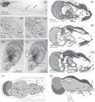
Appendicularians play an important role in marine food webs, but the alimentary canal of hermaphroditic oikopleurids, which prevalent among urochordates, is little studied ultrastructurally. Serial microtomy revealed that the alimentary canal of Oikopleura gracilis consists of a wide pharynx and a U-shaped gut with typical for oikopleurids anatomy: oesophagus, a bilobed stomach, the vertical, short mid- and voluminous distal or rectal intestines, ending the anal papilla. The mid-intestine leaves under the right gastric lobe. TEM analysis diagnosed the following features in the alimentary canal of Oikopleura gracilis. First, the pharynx is lined with a single layer of highly flattened non-ciliated epithelium. Above the anterior part of the endostyle the pharyngeal epithelium forms a thickening – ciliated ring. It is formed by cells of two types: prismatic, ciliated microvillar cells arranged in several parallel rows, and two rows of narrow parietal cells covering the first cell type. Ciliated ring serves as a kind of first valve, prevents food reflux. Second, in the pharynx of one of the specimens three flagellate protozoans with ultrastructurally similarities of free-living kinetoplastids were found. Third, an additional row of gastric band cells was revealed in the upper part of the right gastric lobe compared to that of the gastric floor. Giant electron-lucent vesicles with homogenous content associated with rough endoplasmic reticulum found in the cytoplasm of the gastric band cell, indicating probable storage the protein digestive secrets in these cells. Large multivesicular bodies diagnosed in gastric band cells ultrastructurally corresponded to autophagosomes. Forth, a new cellular type was found, the osmiophilic enterocytes, scattered throughout the digestive epithelium, that shared ultrastructural signs with apoptotic cells. Fifth, TEM data revealed the peritrophic membrane structure and proposed its formation. The absence of its own microflora in the peritrophic membrane, i.e., the absence of an immunity barrier, may be connected with the short ontogeny of appendicularians. The ultrastructural features revealed in the alimentary canal of O. gracilis expand our knowledge of the functioning of the appendicularians and serve as the basis for further research.
期刊介绍:
The Russian Journal of Marine Biology was founded in 1975 by Alexey V. Zhirmunsky, member of the Russian Academy of Sciences. The Russian Journal of Marine Biology covers a wide range of research and some applied aspects of marine biology as a synthetic science related to various fields of study on marine biota and environment. It presents fundamental research on biological processes at molecular, cellular, organismal, and populational levels in marine organisms. Consideration is given to marine objects as models in life sciences. The journal also publishes papers dedicated to events in Russian and international marine biological science and the history of biology.


 分享
分享
 求助内容:
求助内容: 应助结果提醒方式:
 扫码关注我们
