Jeongmin Han, Seong Wook Lee, Na Young Han, Ho-Shin Gwak
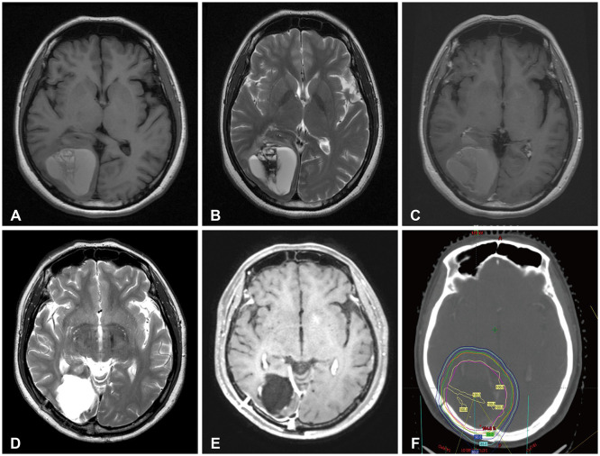
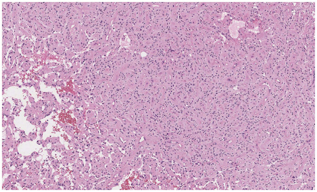
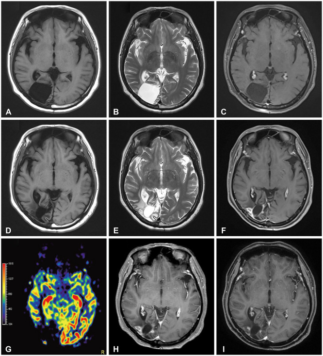
{"title":"Excessively Delayed Radiation Changes After Proton Beam Therapy for Brain Tumors: Report of Two Cases.","authors":"Jeongmin Han, Seong Wook Lee, Na Young Han, Ho-Shin Gwak","doi":"10.14791/btrt.2024.0017","DOIUrl":null,"url":null,"abstract":"<p><p>Delayed cerebral necrosis is a well-known complication of radiation therapy (RT). Because of its irreversible nature, it should be avoided if possible, but avoidance occurs at the expense of potentially compromised tumor control, despite the use of the modern advanced technique of conformal RT that minimizes radiation to normal brain tissue. Risk factors for radiation-induced cerebral necrosis include a higher dose per fraction, larger treatment volume, higher cumulative dose, and shorter time interval (for re-irradiation). The same principle can be applied to proton beam therapy (PBT) to avoid delayed cerebral necrosis. However, conversion of PBT radiation energy into conventional RT is still short of clinical support, compared to conventional RT. Herein, we describe two patients with excessively delayed cerebral necrosis after PBT, in whom follow-up MRI showed no RT-induced changes prior to 3 years after treatment. One patient developed radiation necrosis at 4 years after PBT to the resection cavity of an astroblastoma, and the other developed brainstem necrosis that became symptomatic 6 months after its first appearance on the 3-year follow-up brain MRI. We also discuss possible differences between radiation changes after PBT versus conventional RT.</p>","PeriodicalId":72453,"journal":{"name":"Brain tumor research and treatment","volume":"12 2","pages":"141-147"},"PeriodicalIF":0.0000,"publicationDate":"2024-04-01","publicationTypes":"Journal Article","fieldsOfStudy":null,"isOpenAccess":false,"openAccessPdf":"https://www.ncbi.nlm.nih.gov/pmc/articles/PMC11096628/pdf/","citationCount":"0","resultStr":null,"platform":"Semanticscholar","paperid":null,"PeriodicalName":"Brain tumor research and treatment","FirstCategoryId":"1085","ListUrlMain":"https://doi.org/10.14791/btrt.2024.0017","RegionNum":0,"RegionCategory":null,"ArticlePicture":[],"TitleCN":null,"AbstractTextCN":null,"PMCID":null,"EPubDate":"","PubModel":"","JCR":"","JCRName":"","Score":null,"Total":0}
引用次数: 0
Abstract
Delayed cerebral necrosis is a well-known complication of radiation therapy (RT). Because of its irreversible nature, it should be avoided if possible, but avoidance occurs at the expense of potentially compromised tumor control, despite the use of the modern advanced technique of conformal RT that minimizes radiation to normal brain tissue. Risk factors for radiation-induced cerebral necrosis include a higher dose per fraction, larger treatment volume, higher cumulative dose, and shorter time interval (for re-irradiation). The same principle can be applied to proton beam therapy (PBT) to avoid delayed cerebral necrosis. However, conversion of PBT radiation energy into conventional RT is still short of clinical support, compared to conventional RT. Herein, we describe two patients with excessively delayed cerebral necrosis after PBT, in whom follow-up MRI showed no RT-induced changes prior to 3 years after treatment. One patient developed radiation necrosis at 4 years after PBT to the resection cavity of an astroblastoma, and the other developed brainstem necrosis that became symptomatic 6 months after its first appearance on the 3-year follow-up brain MRI. We also discuss possible differences between radiation changes after PBT versus conventional RT.


 分享
分享

 求助内容:
求助内容: 应助结果提醒方式:
 扫码关注我们
