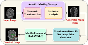
{"title":"Enhancing clinical diagnostics: novel denoising methodology for brain MRI with adaptive masking and modified non-local block.","authors":"A Velayudham, K Madhan Kumar, Krishna Priya M S","doi":"10.1007/s11517-024-03122-y","DOIUrl":null,"url":null,"abstract":"<p><p>Medical image denoising has been a subject of extensive research, with various techniques employed to enhance image quality and facilitate more accurate diagnostics. The evolution of denoising methods has highlighted impressive results but struggled to strike equilibrium between noise reduction and edge preservation which limits its applicability in various domains. This paper manifests the novel methodology that integrates an adaptive masking strategy, transformer-based U-Net Prior generator, edge enhancement module, and modified non-local block (MNLB) for denoising brain MRI clinical images. The adaptive masking strategy maintains the vital information through dynamic mask generation while the prior generator by capturing hierarchical features regenerates the high-quality prior MRI images. Finally, these images are fed to the edge enhancement module to boost structural information by maintaining crucial edge details, and the MNLB produces the denoised output by deriving non-local contextual information. The comprehensive experimental assessment is performed by employing two datasets namely the brain tumor MRI dataset and Alzheimer's dataset for diverse metrics and compared with conventional denoising approaches. The proposed denoising methodology achieves a PSNR of 40.965 and SSIM of 0.938 on the Alzheimer's dataset and also achieves a PSNR of 40.002 and SSIM of 0.926 on the brain tumor MRI dataset at a noise level of 50% revealing its supremacy in noise minimization. Furthermore, the impact of different masking ratios on denoising performance is analyzed which reveals that the proposed method showed PSNR of 40.965, SSIM of 0.938, MAE of 5.847, and MSE of 3.672 at the masking ratio of 60%. Moreover, the findings pave the way for the advancement of clinical image processing, facilitating precise detection of tumors in clinical MRI images.</p>","PeriodicalId":49840,"journal":{"name":"Medical & Biological Engineering & Computing","volume":" ","pages":"3043-3056"},"PeriodicalIF":2.6000,"publicationDate":"2024-10-01","publicationTypes":"Journal Article","fieldsOfStudy":null,"isOpenAccess":false,"openAccessPdf":"","citationCount":"0","resultStr":null,"platform":"Semanticscholar","paperid":null,"PeriodicalName":"Medical & Biological Engineering & Computing","FirstCategoryId":"5","ListUrlMain":"https://doi.org/10.1007/s11517-024-03122-y","RegionNum":4,"RegionCategory":"医学","ArticlePicture":[],"TitleCN":null,"AbstractTextCN":null,"PMCID":null,"EPubDate":"2024/5/18 0:00:00","PubModel":"Epub","JCR":"Q2","JCRName":"COMPUTER SCIENCE, INTERDISCIPLINARY APPLICATIONS","Score":null,"Total":0}
引用次数: 0
Abstract
Medical image denoising has been a subject of extensive research, with various techniques employed to enhance image quality and facilitate more accurate diagnostics. The evolution of denoising methods has highlighted impressive results but struggled to strike equilibrium between noise reduction and edge preservation which limits its applicability in various domains. This paper manifests the novel methodology that integrates an adaptive masking strategy, transformer-based U-Net Prior generator, edge enhancement module, and modified non-local block (MNLB) for denoising brain MRI clinical images. The adaptive masking strategy maintains the vital information through dynamic mask generation while the prior generator by capturing hierarchical features regenerates the high-quality prior MRI images. Finally, these images are fed to the edge enhancement module to boost structural information by maintaining crucial edge details, and the MNLB produces the denoised output by deriving non-local contextual information. The comprehensive experimental assessment is performed by employing two datasets namely the brain tumor MRI dataset and Alzheimer's dataset for diverse metrics and compared with conventional denoising approaches. The proposed denoising methodology achieves a PSNR of 40.965 and SSIM of 0.938 on the Alzheimer's dataset and also achieves a PSNR of 40.002 and SSIM of 0.926 on the brain tumor MRI dataset at a noise level of 50% revealing its supremacy in noise minimization. Furthermore, the impact of different masking ratios on denoising performance is analyzed which reveals that the proposed method showed PSNR of 40.965, SSIM of 0.938, MAE of 5.847, and MSE of 3.672 at the masking ratio of 60%. Moreover, the findings pave the way for the advancement of clinical image processing, facilitating precise detection of tumors in clinical MRI images.
期刊介绍:
Founded in 1963, Medical & Biological Engineering & Computing (MBEC) continues to serve the biomedical engineering community, covering the entire spectrum of biomedical and clinical engineering. The journal presents exciting and vital experimental and theoretical developments in biomedical science and technology, and reports on advances in computer-based methodologies in these multidisciplinary subjects. The journal also incorporates new and evolving technologies including cellular engineering and molecular imaging.
MBEC publishes original research articles as well as reviews and technical notes. Its Rapid Communications category focuses on material of immediate value to the readership, while the Controversies section provides a forum to exchange views on selected issues, stimulating a vigorous and informed debate in this exciting and high profile field.
MBEC is an official journal of the International Federation of Medical and Biological Engineering (IFMBE).


 分享
分享
 求助内容:
求助内容: 应助结果提醒方式:
 扫码关注我们
