{"title":"An analysis of semaphorin-mediated cellular interactions in the Caenorhabditis elegans epidermis using the IR-LEGO single-cell gene induction system","authors":"Motoshi Suzuki, Shin Takagi","doi":"10.1111/dgd.12925","DOIUrl":null,"url":null,"abstract":"<p>One of the major functions of the semaphorin signaling system is the regulation of cell shape. In the nematode <i>Caenorhabditis elegans</i>, membrane-bound semaphorins SMP-1/2 (SMPs) regulate the morphology of epidermal cells via their receptor plexin, PLX-1. In the larval male tail of the SMP-PLX-1 signaling mutants, the border between two epidermal cells, R1.p and R2.p, is displaced anteriorly, resulting in the anterior displacement of the anterior-most ray, ray 1, in the adult male. To elucidate how the intercellular signaling mediated by SMPs regulates the position of the intercellular border, we performed mosaic gene expression analyses by using infrared laser-evoked gene operator (IR-LEGO). We show that PLX-1 expressed in R1.p and SMP-1 expressed in R2.p are required for the proper positioning of ray 1. The result suggests that SMP signaling promotes extension, rather than retraction, of R1.p. This is in contrast to a previous finding that SMPs mediate inhibition of cell extension of vulval precursor cells, another group of epidermal cells of <i>C. elegans</i>, indicating the context dependence of cell shape control via the semaphorin signaling system.</p>","PeriodicalId":50589,"journal":{"name":"Development Growth & Differentiation","volume":"66 5","pages":"308-319"},"PeriodicalIF":1.0000,"publicationDate":"2024-05-18","publicationTypes":"Journal Article","fieldsOfStudy":null,"isOpenAccess":false,"openAccessPdf":"https://onlinelibrary.wiley.com/doi/epdf/10.1111/dgd.12925","citationCount":"0","resultStr":null,"platform":"Semanticscholar","paperid":null,"PeriodicalName":"Development Growth & Differentiation","FirstCategoryId":"99","ListUrlMain":"https://onlinelibrary.wiley.com/doi/10.1111/dgd.12925","RegionNum":4,"RegionCategory":"生物学","ArticlePicture":[],"TitleCN":null,"AbstractTextCN":null,"PMCID":null,"EPubDate":"","PubModel":"","JCR":"Q4","JCRName":"CELL BIOLOGY","Score":null,"Total":0}
引用次数: 0
Abstract
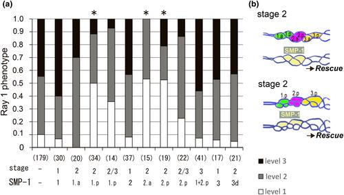
One of the major functions of the semaphorin signaling system is the regulation of cell shape. In the nematode Caenorhabditis elegans, membrane-bound semaphorins SMP-1/2 (SMPs) regulate the morphology of epidermal cells via their receptor plexin, PLX-1. In the larval male tail of the SMP-PLX-1 signaling mutants, the border between two epidermal cells, R1.p and R2.p, is displaced anteriorly, resulting in the anterior displacement of the anterior-most ray, ray 1, in the adult male. To elucidate how the intercellular signaling mediated by SMPs regulates the position of the intercellular border, we performed mosaic gene expression analyses by using infrared laser-evoked gene operator (IR-LEGO). We show that PLX-1 expressed in R1.p and SMP-1 expressed in R2.p are required for the proper positioning of ray 1. The result suggests that SMP signaling promotes extension, rather than retraction, of R1.p. This is in contrast to a previous finding that SMPs mediate inhibition of cell extension of vulval precursor cells, another group of epidermal cells of C. elegans, indicating the context dependence of cell shape control via the semaphorin signaling system.
期刊介绍:
Development Growth & Differentiation (DGD) publishes three types of articles: original, resource, and review papers.
Original papers are on any subjects having a context in development, growth, and differentiation processes in animals, plants, and microorganisms, dealing with molecular, genetic, cellular and organismal phenomena including metamorphosis and regeneration, while using experimental, theoretical, and bioinformatic approaches. Papers on other related fields are also welcome, such as stem cell biology, genomics, neuroscience, Evodevo, Ecodevo, and medical science as well as related methodology (new or revised techniques) and bioresources.
Resource papers describe a dataset, such as whole genome sequences and expressed sequence tags (ESTs), with some biological insights, which should be valuable for studying the subjects as mentioned above.
Submission of review papers is also encouraged, especially those providing a new scope based on the authors’ own study, or a summarization of their study series.


 分享
分享
 求助内容:
求助内容: 应助结果提醒方式:
 扫码关注我们
