Wojciech Baran, Zdzisław Woźniak, Aleksandra Batycka-Baran
{"title":"IL-17: a novel player in the pathogenesis of vulvar lichen sclerosus.","authors":"Wojciech Baran, Zdzisław Woźniak, Aleksandra Batycka-Baran","doi":"10.5114/ada.2024.139142","DOIUrl":null,"url":null,"abstract":"<p><strong>Introduction: </strong>Vulvar lichen sclerosus (VLS) is a chronic progressive, lymphocyte-mediated inflammatory disease whose pathogenesis is complex and not fully elucidated.</p><p><strong>Aim: </strong>In the current study we have investigated for the first time the expression of interleukin-17 (IL-17) and S100A7 in lesional skin obtained from female individuals with histologically confirmed VLS.</p><p><strong>Material and methods: </strong>In our study we used skin biopsies obtained from female patients with histologically confirmed VLS (<i>n</i> = 20) and skin samples from healthy age- and gender-matched individuals (plastic surgery procedures) (<i>n</i> = 10) serving as controls. The tissue expressions of IL-17 and S100A7 were assessed with an immunohistochemical method.</p><p><strong>Results: </strong>The number of cells showing IL-17 expression was significantly higher in VLS lesional skin as compared to normal skin of healthy controls (<i>p</i> < 0.0001). In VLS lesional skin, IL-17 was expressed in the epidermis and by cells within the inflammatory infiltrate in the upper dermis. The number of cells showing S100A7 expression was significantly higher in VLS lesional skin as compared to normal skin of healthy controls (<i>p</i> < 0.0001). In VLS lesional skin, S100A7 was expressed by suprabasal keratinocytes in epidermis. S100A7 was also expressed by cells within the inflammatory infiltrate in the dermis.</p><p><strong>Conclusions: </strong>The results of our study may suggest the involvement of IL-17 and S100A7 in the pathogenesis of VLS. The better understanding of this disease may lead to the development of novel, effective therapeutic strategies e.g. using well-known biologics IL-17 inhibitors class.</p>","PeriodicalId":54595,"journal":{"name":"Postepy Dermatologii I Alergologii","volume":"41 2","pages":"220-225"},"PeriodicalIF":1.4000,"publicationDate":"2024-04-01","publicationTypes":"Journal Article","fieldsOfStudy":null,"isOpenAccess":false,"openAccessPdf":"https://www.ncbi.nlm.nih.gov/pmc/articles/PMC11110226/pdf/","citationCount":"0","resultStr":null,"platform":"Semanticscholar","paperid":null,"PeriodicalName":"Postepy Dermatologii I Alergologii","FirstCategoryId":"3","ListUrlMain":"https://doi.org/10.5114/ada.2024.139142","RegionNum":4,"RegionCategory":"医学","ArticlePicture":[],"TitleCN":null,"AbstractTextCN":null,"PMCID":null,"EPubDate":"2024/4/24 0:00:00","PubModel":"Epub","JCR":"Q3","JCRName":"ALLERGY","Score":null,"Total":0}
引用次数: 0
Abstract
Introduction: Vulvar lichen sclerosus (VLS) is a chronic progressive, lymphocyte-mediated inflammatory disease whose pathogenesis is complex and not fully elucidated.
Aim: In the current study we have investigated for the first time the expression of interleukin-17 (IL-17) and S100A7 in lesional skin obtained from female individuals with histologically confirmed VLS.
Material and methods: In our study we used skin biopsies obtained from female patients with histologically confirmed VLS (n = 20) and skin samples from healthy age- and gender-matched individuals (plastic surgery procedures) (n = 10) serving as controls. The tissue expressions of IL-17 and S100A7 were assessed with an immunohistochemical method.
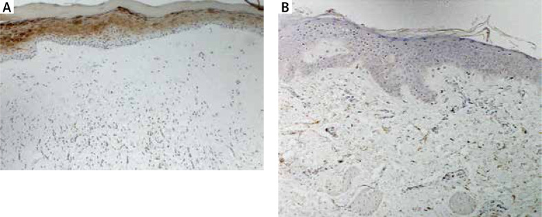
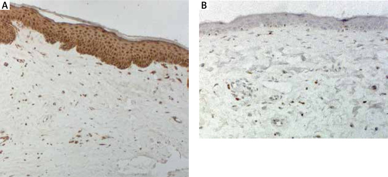
Results: The number of cells showing IL-17 expression was significantly higher in VLS lesional skin as compared to normal skin of healthy controls (p < 0.0001). In VLS lesional skin, IL-17 was expressed in the epidermis and by cells within the inflammatory infiltrate in the upper dermis. The number of cells showing S100A7 expression was significantly higher in VLS lesional skin as compared to normal skin of healthy controls (p < 0.0001). In VLS lesional skin, S100A7 was expressed by suprabasal keratinocytes in epidermis. S100A7 was also expressed by cells within the inflammatory infiltrate in the dermis.
Conclusions: The results of our study may suggest the involvement of IL-17 and S100A7 in the pathogenesis of VLS. The better understanding of this disease may lead to the development of novel, effective therapeutic strategies e.g. using well-known biologics IL-17 inhibitors class.


 分享
分享
 求助内容:
求助内容: 应助结果提醒方式:
 扫码关注我们
