Lea Christierson, Petter Frieberg, Tania Lala, Johannes Töger, Petru Liuba, Johan Revstedt, Hanna Isaksson, Nina Hakacova
{"title":"Multi-Modal in Vitro Experiments Mimicking the Flow Through a Mitral Heart Valve Phantom.","authors":"Lea Christierson, Petter Frieberg, Tania Lala, Johannes Töger, Petru Liuba, Johan Revstedt, Hanna Isaksson, Nina Hakacova","doi":"10.1007/s13239-024-00732-3","DOIUrl":null,"url":null,"abstract":"<p><strong>Purpose: </strong>Fluid-structure interaction (FSI) models are more commonly applied in medical research as computational power is increasing. However, understanding the accuracy of FSI models is crucial, especially in the context of heart valve disease in patient-specific models. Therefore, this study aimed to create a multi-modal benchmarking data set for cardiac-inspired FSI models, based on clinically important parameters, such as the pressure, velocity, and valve opening, with an in vitro phantom setup.</p><p><strong>Method: </strong>An in vitro setup was developed with a 3D-printed phantom mimicking the left heart, including a deforming mitral valve. A range of pulsatile flows were created with a computer-controlled motor-and-pump setup. Catheter pressure measurements, magnetic resonance imaging (MRI), and echocardiography (Echo) imaging were used to measure pressure and velocity in the domain. Furthermore, the valve opening was quantified based on cine MRI and Echo images.</p><p><strong>Result: </strong>The experimental setup, with 0.5% cycle-to-cycle variation, was successfully built and six different flow cases were investigated. Higher velocity through the mitral valve was observed for increased cardiac output. The pressure difference across the valve also followed this trend. The flow in the phantom was qualitatively assessed by the velocity profile in the ventricle and by streamlines obtained from 4D phase-contrast MRI.</p><p><strong>Conclusion: </strong>A multi-modal set of data for validation of FSI models has been created, based on parameters relevant for diagnosis of heart valve disease. All data is publicly available for future development of computational heart valve models.</p>","PeriodicalId":54322,"journal":{"name":"Cardiovascular Engineering and Technology","volume":" ","pages":"572-583"},"PeriodicalIF":1.8000,"publicationDate":"2024-10-01","publicationTypes":"Journal Article","fieldsOfStudy":null,"isOpenAccess":false,"openAccessPdf":"https://www.ncbi.nlm.nih.gov/pmc/articles/PMC11582118/pdf/","citationCount":"0","resultStr":null,"platform":"Semanticscholar","paperid":null,"PeriodicalName":"Cardiovascular Engineering and Technology","FirstCategoryId":"5","ListUrlMain":"https://doi.org/10.1007/s13239-024-00732-3","RegionNum":4,"RegionCategory":"医学","ArticlePicture":[],"TitleCN":null,"AbstractTextCN":null,"PMCID":null,"EPubDate":"2024/5/23 0:00:00","PubModel":"Epub","JCR":"Q3","JCRName":"CARDIAC & CARDIOVASCULAR SYSTEMS","Score":null,"Total":0}
引用次数: 0
Abstract
Purpose: Fluid-structure interaction (FSI) models are more commonly applied in medical research as computational power is increasing. However, understanding the accuracy of FSI models is crucial, especially in the context of heart valve disease in patient-specific models. Therefore, this study aimed to create a multi-modal benchmarking data set for cardiac-inspired FSI models, based on clinically important parameters, such as the pressure, velocity, and valve opening, with an in vitro phantom setup.
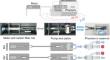
Method: An in vitro setup was developed with a 3D-printed phantom mimicking the left heart, including a deforming mitral valve. A range of pulsatile flows were created with a computer-controlled motor-and-pump setup. Catheter pressure measurements, magnetic resonance imaging (MRI), and echocardiography (Echo) imaging were used to measure pressure and velocity in the domain. Furthermore, the valve opening was quantified based on cine MRI and Echo images.
Result: The experimental setup, with 0.5% cycle-to-cycle variation, was successfully built and six different flow cases were investigated. Higher velocity through the mitral valve was observed for increased cardiac output. The pressure difference across the valve also followed this trend. The flow in the phantom was qualitatively assessed by the velocity profile in the ventricle and by streamlines obtained from 4D phase-contrast MRI.
Conclusion: A multi-modal set of data for validation of FSI models has been created, based on parameters relevant for diagnosis of heart valve disease. All data is publicly available for future development of computational heart valve models.
期刊介绍:
Cardiovascular Engineering and Technology is a journal publishing the spectrum of basic to translational research in all aspects of cardiovascular physiology and medical treatment. It is the forum for academic and industrial investigators to disseminate research that utilizes engineering principles and methods to advance fundamental knowledge and technological solutions related to the cardiovascular system. Manuscripts spanning from subcellular to systems level topics are invited, including but not limited to implantable medical devices, hemodynamics and tissue biomechanics, functional imaging, surgical devices, electrophysiology, tissue engineering and regenerative medicine, diagnostic instruments, transport and delivery of biologics, and sensors. In addition to manuscripts describing the original publication of research, manuscripts reviewing developments in these topics or their state-of-art are also invited.


 分享
分享
 求助内容:
求助内容: 应助结果提醒方式:
 扫码关注我们
